File:Phase-contrast images of embryos at different developmental stages via neogenin expression.png
Original file (1,938 × 2,171 pixels, file size: 4.85 MB, MIME type: image/png)
Phase-contrast images of embryos at different developmental stages via neogenin expression
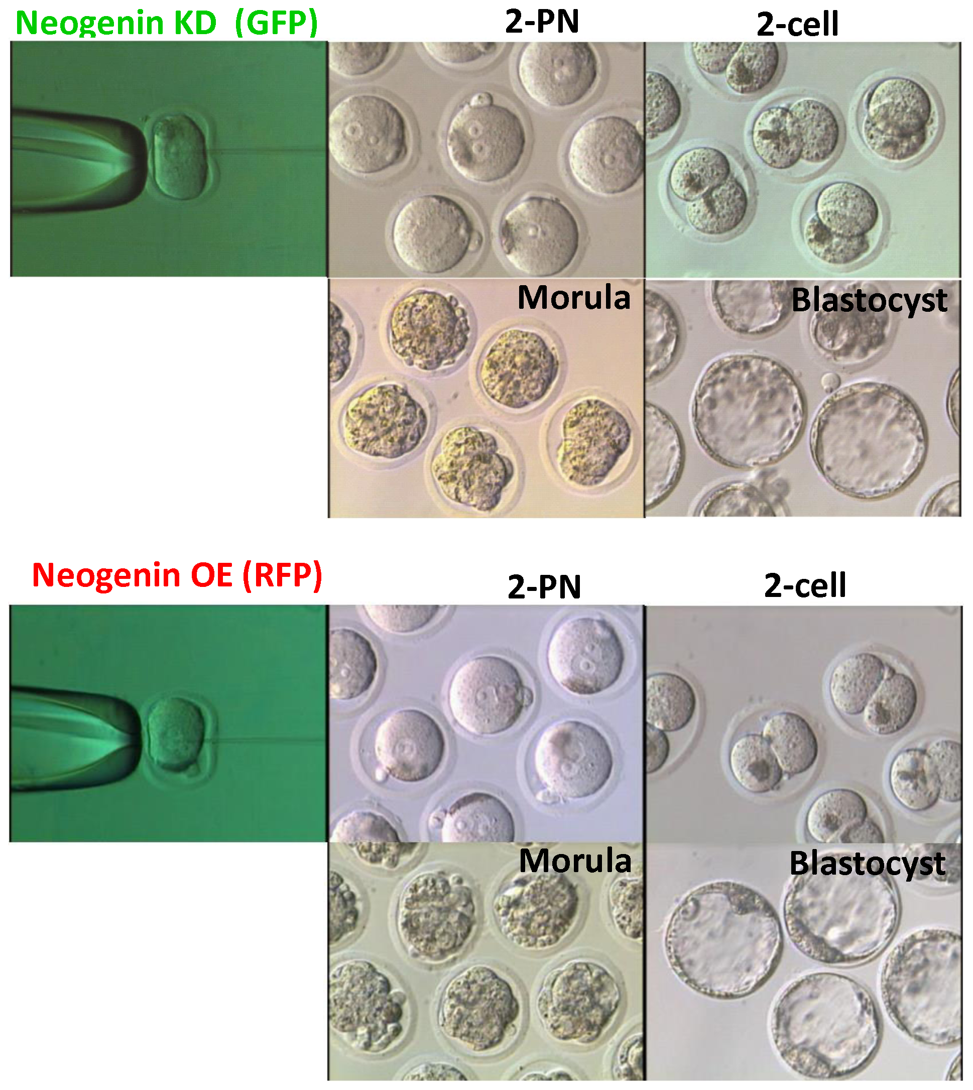
FIGURE 2: Effects of up-regulation and down-regulation of neogenin on embryo development. 2-PN mouse zygotes were microinjected with shRNA targeting neogenin (neogenin KD) or with neogenin cDNA vectors (neogenin OE) and were cultured to the blastocyst stage. To visually differentiate neogenin KD from neogenin OE embryos, GFP and RFP were co-expressed, respectively.Phase-contrast images of embryos at different developmental stages were observed.
--Mark Hill (talk) 16:15, 21 August 2014 (EST) This is all correct. The image is very large (4.8 MB), perhaps a small version could have been uploaded. You can adjust the resolution and size in most image editing programs.
Reference
<pubmed>25013897</pubmed>| PLos One
Copyright
© 2014 Lee et al. This is an open-access article distributed under the terms of the Creative Commons Attribution License, which permits unrestricted use, distribution, and reproduction in any medium, provided the original author and source are credited.
- Note - This image was originally uploaded as part of an undergraduate science student project and may contain inaccuracies in either description or acknowledgements. Students have been advised in writing concerning the reuse of content and may accidentally have misunderstood the original terms of use. If image reuse on this non-commercial educational site infringes your existing copyright, please contact the site editor for immediate removal.
File history
Click on a date/time to view the file as it appeared at that time.
| Date/Time | Thumbnail | Dimensions | User | Comment | |
|---|---|---|---|---|---|
| current | 03:31, 20 August 2014 | | 1,938 × 2,171 (4.85 MB) | Z3418837 (talk | contribs) |
You cannot overwrite this file.
File usage
The following page uses this file: