File:Pharyngeal arch cartilages.jpg
Pharyngeal_arch_cartilages.jpg (400 × 324 pixels, file size: 26 KB, MIME type: image/jpeg)
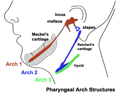
Pharyngeal Arch Cartilages
Meckel's cartilage - located within the first pharyngeal arch mandibular prominence, forms a cartilage "template" besides which the mandible develops by the process of intramembranous ossification. It is important to note that this cartilage template does not ossify (endochondral ossification) but provides a transient structure where the mandible will form, and later degenerates. The ends of this "U" shaped cartilage form two of the middle ear ossicles (malleous and incus).
Reichert's cartilage - located within the second pharyngeal arch. The ends of this "U" shaped cartilage form the stapes of the middle ear ossicles.
- Links: Middle Ear | Skull Development | Head Development
Source: UNSW Embryology
File history
Click on a date/time to view the file as it appeared at that time.
| Date/Time | Thumbnail | Dimensions | User | Comment | |
|---|---|---|---|---|---|
| current | 23:41, 13 May 2009 | | 400 × 324 (26 KB) | MarkHill (talk | contribs) | Pharyngeal Arch cartilages Source: UNSW Embryology |
You cannot overwrite this file.
File usage
The following 23 pages use this file:
- 2009 BGD-B Lecture Face and Ear
- 2009 Lecture 11
- 2009 Lecture 17
- 2010 Lecture 11
- 2010 Lecture 17
- 2011 Lab 10 - Early Embryo
- 2011 Lab 6 - Early Embryo
- AACP Meeting 2013 - Face Embryology
- ANAT2341 Lab 10 - Early Embryo
- ANAT2341 Lab 6 - Early Embryo
- Abnormal Development - Cleft Lip and Palate
- BGDB Face and Ear - Early Embryo
- BGD Lecture - Face and Ear Development
- Head Development
- Hearing - Middle Ear Development
- Lecture - Head Development
- Lecture - Sensory Development
- Musculoskeletal System - Skull Development
- Neural Crest - Cranial Nerve Development
- Neural Crest - Cranial Nerves
- Neural Crest Development
- Palate Development
- R