File:Pharyngeal arch cartilages.jpg
Pharyngeal_arch_cartilages.jpg (400 × 324 pixels, file size: 26 KB, MIME type: image/jpeg)
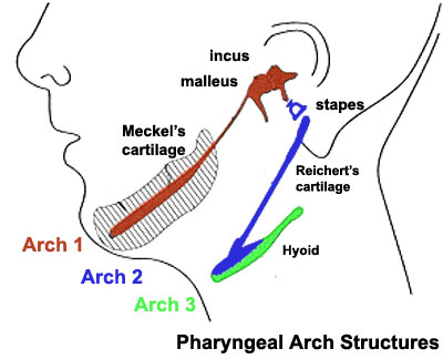
Pharyngeal Arch Cartilages
Meckel's cartilage - located within the first pharyngeal arch mandibular prominence, forms a cartilage "template" besides which the mandible develops by the process of intramembranous ossification. It is important to note that this cartilage template does not ossify (endochondral ossification) but provides a transient structure where the mandible will form, and later degenerates. The ends of this "U" shaped cartilage form two of the middle ear ossicles (malleous and incus).
Reichert's cartilage - located within the second pharyngeal arch. The ventral portion contributes the superior part of the hyoid cartilage. The ends of this "U" shaped cartilage form the stapes of the middle ear ossicles.
- Links: Middle Ear | Skull Development | Head Development[[Category:
File history
Click on a date/time to view the file as it appeared at that time.
| Date/Time | Thumbnail | Dimensions | User | Comment | |
|---|---|---|---|---|---|
| current | 23:41, 13 May 2009 | | 400 × 324 (26 KB) | MarkHill (talk | contribs) | Pharyngeal Arch cartilages Source: UNSW Embryology |
You cannot overwrite this file.
File usage
The following 23 pages use this file:
- 2009 BGD-B Lecture Face and Ear
- 2009 Lecture 11
- 2009 Lecture 17
- 2010 Lecture 11
- 2010 Lecture 17
- 2011 Lab 10 - Early Embryo
- 2011 Lab 6 - Early Embryo
- AACP Meeting 2013 - Face Embryology
- ANAT2341 Lab 10 - Early Embryo
- ANAT2341 Lab 6 - Early Embryo
- Abnormal Development - Cleft Lip and Palate
- BGDB Face and Ear - Early Embryo
- BGD Lecture - Face and Ear Development
- Head Development
- Hearing - Middle Ear Development
- Lecture - Head Development
- Lecture - Sensory Development
- Musculoskeletal System - Skull Development
- Neural Crest - Cranial Nerve Development
- Neural Crest - Cranial Nerves
- Neural Crest Development
- Palate Development
- R