File:Peters1899 plate14.jpg

Original file (2,000 × 2,527 pixels, file size: 343 KB, MIME type: image/jpeg)
Plate 14.
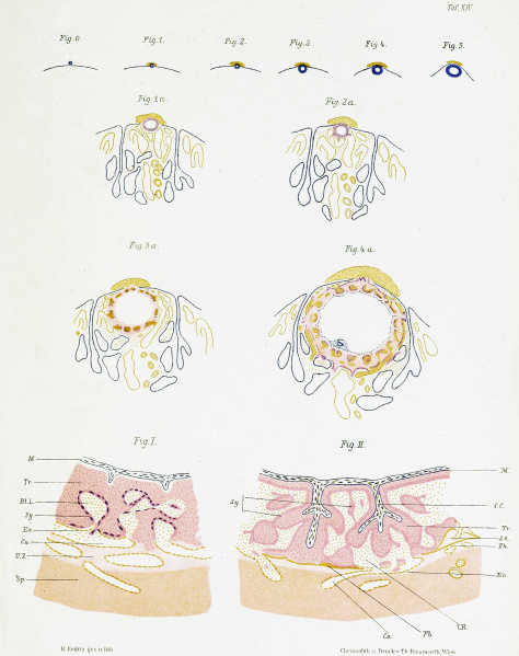
Fig. 0 bis 5 schematische Darstellung der Einbettung des menschlichen Eies in annähernd natürlichen Dimensionen.
Fig. 1 a bis 4 a. Obige Figuren 1 bis 4 grosser dargestellt.
Fig. I und Fig. II. Schemata der Placentarentwickelung.
M. Mesoderm, Tr. Trophoblast, BLL. Blutlacunen, i.R intravillöser Raum, Sy, Syneytium, En. Endothel, Ca. mṻtterliche Capillaren, U.Z. Umlagerungszone, Sp. Spongiosa, f.C. fotale Capillaren, d.S. deciduales Septum, Fb. Fibrin.
Reference
Peters H. Ueber die Einbettung des menschlichen Eies und das früheste, bisher bekannte, menschliche Placentationsstadium (About the embedding of the human egg and the earliest known human placentation stage). (1899) Deuticke, Leipzig.
Cite this page: Hill, M.A. (2024, April 27) Embryology Peters1899 plate14.jpg. Retrieved from https://embryology.med.unsw.edu.au/embryology/index.php/File:Peters1899_plate14.jpg
- © Dr Mark Hill 2024, UNSW Embryology ISBN: 978 0 7334 2609 4 - UNSW CRICOS Provider Code No. 00098G
File history
Click on a date/time to view the file as it appeared at that time.
| Date/Time | Thumbnail | Dimensions | User | Comment | |
|---|---|---|---|---|---|
| current | 14:57, 21 August 2018 |  | 2,000 × 2,527 (343 KB) | Z8600021 (talk | contribs) | |
| 14:46, 21 August 2018 |  | 2,330 × 2,944 (419 KB) | Z8600021 (talk | contribs) |
You cannot overwrite this file.
File usage
The following page uses this file: