File:Pearce1903 fig01.jpg
From Embryology
Size of this preview: 667 × 600 pixels. Other resolution: 900 × 809 pixels.
Original file (900 × 809 pixels, file size: 335 KB, MIME type: image/jpeg)
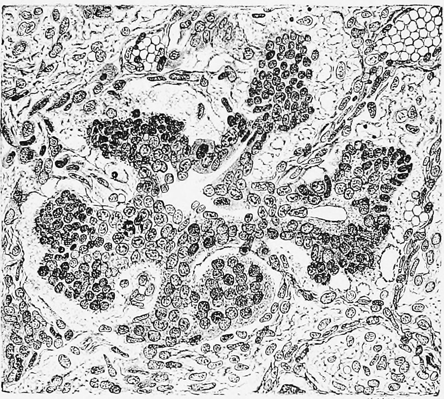
Fig. 1. 1. Pancreas of an embryo 54 mm in length
Early stage of the differentiation of an island or pond to those described by Langerhans. A round mass composed of cells rich In protoplasm is seen continuous with the guesse and Renaut, but I have periphery of the gland.
Reference
Pearce RM. The development of the islands of Langerhans in the human embryo. (1903) Amer. J Anat. : 446-455.
Cite this page: Hill, M.A. (2024, June 1) Embryology Pearce1903 fig01.jpg. Retrieved from https://embryology.med.unsw.edu.au/embryology/index.php/File:Pearce1903_fig01.jpg
- © Dr Mark Hill 2024, UNSW Embryology ISBN: 978 0 7334 2609 4 - UNSW CRICOS Provider Code No. 00098G
File history
Click on a date/time to view the file as it appeared at that time.
| Date/Time | Thumbnail | Dimensions | User | Comment | |
|---|---|---|---|---|---|
| current | 15:23, 2 August 2019 | | 900 × 809 (335 KB) | Z8600021 (talk | contribs) | |
| 15:19, 2 August 2019 |  | 978 × 1,109 (319 KB) | Z8600021 (talk | contribs) | {{Ref-Pearce1903}} |
You cannot overwrite this file.
File usage
The following page uses this file: