File:Patten1938 text-fig02.jpg
Original file (1,000 × 839 pixels, file size: 173 KB, MIME type: image/jpeg)
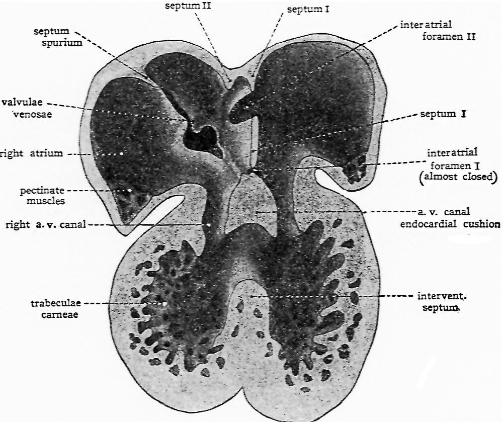
Text-Fig. 2. Semi-schematic drawing of the interior of the heart
To show the start of interatrial septum sectmdum and the appearance of interatrial foramen secundum in septum primum. Based on original reconstructions of the heart of a 9.4 mm pig embryo and on Tandler’s reconstructions of the heart of human embryos of the 7th week. (From Embryology, Patten, B. M., courtesy P. Blakiston’s Son & Co.)
Reference
Patten BM. Developmental defects at the foramen ovale. (1938) Am J Pathol. 14(2):135-162. PMID 19970381
Cite this page: Hill, M.A. (2024, April 28) Embryology Patten1938 text-fig02.jpg. Retrieved from https://embryology.med.unsw.edu.au/embryology/index.php/File:Patten1938_text-fig02.jpg
- © Dr Mark Hill 2024, UNSW Embryology ISBN: 978 0 7334 2609 4 - UNSW CRICOS Provider Code No. 00098G
File history
Click on a date/time to view the file as it appeared at that time.
| Date/Time | Thumbnail | Dimensions | User | Comment | |
|---|---|---|---|---|---|
| current | 15:53, 27 February 2017 | | 1,000 × 839 (173 KB) | Z8600021 (talk | contribs) | |
| 15:53, 27 February 2017 |  | 1,279 × 1,335 (290 KB) | Z8600021 (talk | contribs) | Text-Fig. 2. Semi-schematic drawing of the interior of the heart to show the start of interatrial septum sectmdum and the appearance of interatrial foramen secundum in septum primum. Based on original reconstructions of the heart of a 9.4 mm. pig embry... |
You cannot overwrite this file.
File usage
The following page uses this file: