File:Patten029.jpg
Original file (685 × 676 pixels, file size: 101 KB, MIME type: image/jpeg)
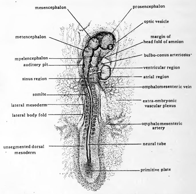
Fig. 29. Dorsal view of entire chick embryo having 19 pairs of somites
(x14) (about 43 hours incubation).
Due to torsion the cephalic region appears in dextro-dorsal view.
The vessels destined to carry blood from the embryo to the vitelline plexus develop in embryos of about 40 hours (Fig. 29). Like the afferent vitelline channels, the efferent channels have a dual origin. The proximal portions of the efferent channels arise within the embryo as branches of the dorsal aortae, and extend peripherally. The distal portions of the channels arise in the extra-embryonic vascular area and extend toward the embryo. The efferent vitelline vessels are estabhshed when these two sets of channels become confluent. In its early stages the connection is through a network of small channels rather than definite vessels, the aortae breaking up posteriorly into small channels some of which communicate laterally with the extra-embryonic plexus. Later some of these channels become confluent, others disappear, and gradually definite main vessels, the omphalomesenteric arteries, are established. For some time after their formation, the omphalomesenteric arteries are likely to retain traces of their origin from a plexus of small channels and arise from the aorta by several roots (Fig. 35).
- Links: Introduction | Gametes and Fertilization | Segmentation | Entoderm | Primitive Streak and Mesoderm | Primitive Streak to Somites | 24 Hours | 24 to 33 Hours | 33 to 39 Hours | 40 to 50 Hours | Extra-embryonic Membranes | 50 to 55 Hours | Day 3 to 4 | References | All Figures
| Historic Disclaimer - information about historic embryology pages |
|---|
 |
Reference
Patten BM. The Early Embryology of the Chick. (1920) Philadelphia: P. Blakiston's Son and Co.
Cite this page: Hill, M.A. (2024, April 26) Embryology Patten029.jpg. Retrieved from https://embryology.med.unsw.edu.au/embryology/index.php/File:Patten029.jpg
- © Dr Mark Hill 2024, UNSW Embryology ISBN: 978 0 7334 2609 4 - UNSW CRICOS Provider Code No. 00098G
File history
Click on a date/time to view the file as it appeared at that time.
| Date/Time | Thumbnail | Dimensions | User | Comment | |
|---|---|---|---|---|---|
| current | 01:59, 17 January 2011 | | 685 × 676 (101 KB) | S8600021 (talk | contribs) | {{Template:Patten_1920_Figures}} |
You cannot overwrite this file.
File usage
The following 2 pages use this file: