File:Ovary- histology overview.jpg
From Embryology
Size of this preview: 799 × 599 pixels. Other resolution: 861 × 646 pixels.
Original file (861 × 646 pixels, file size: 160 KB, MIME type: image/jpeg)
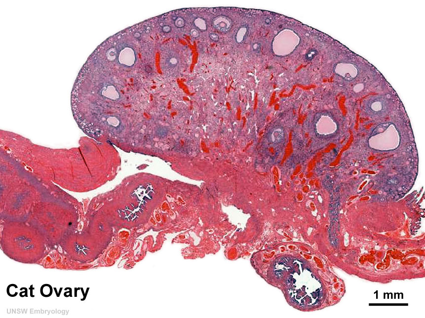
Ovary Histology overview (cat)
Cross-section of the ovary and adjacent oviduct of a cat. At this magnification, the overall organization of the ovary can be observed, cortex/medulla organization and arrangement of the maternal blood vessels, but few specific follicle details can be seen.
Image (low magnification) showing cortical primordial follicles with primary (preantral) and secondary (antral) follicles lying deeper.
Mesovarium at lower right and blood vessels in medullary region.
- Links: Ovary Development | Oocyte Development
File history
Click on a date/time to view the file as it appeared at that time.
| Date/Time | Thumbnail | Dimensions | User | Comment | |
|---|---|---|---|---|---|
| current | 10:58, 9 May 2010 | | 861 × 646 (160 KB) | S8600021 (talk | contribs) | Ovary- histology overview (cat) |
You cannot overwrite this file.
File usage
The following 5 pages use this file: