File:Ossification endochondral 01.jpg
From Embryology
Size of this preview: 799 × 599 pixels. Other resolution: 817 × 613 pixels.
Original file (817 × 613 pixels, file size: 198 KB, MIME type: image/jpeg)
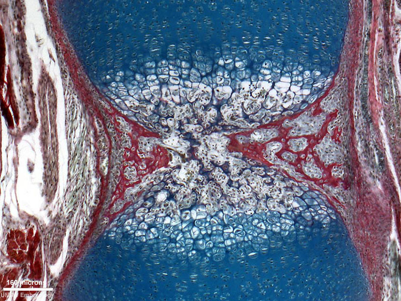
Developing Vertebra - Endochondral Ossification
- Histological image of a developing vertebra (neonatal rat).
- vertebra - cartilage template and developing bony collar (centre of image)
Legend
- blue - cartilage matrix
- red - bone matrix
scale bar 160 microns
Original File Name: Endochondral9x5-1000px.jpg
Image Source: UNSW Embryology
File history
Click on a date/time to view the file as it appeared at that time.
| Date/Time | Thumbnail | Dimensions | User | Comment | |
|---|---|---|---|---|---|
| current | 12:33, 23 March 2012 | | 817 × 613 (198 KB) | Z8600021 (talk | contribs) | ==Developing Vertebra - Endochondral Ossification== * Histological image of a developing vertebra and intervertebral disc (neonatal rat). * intervertebral disc - nucleus pulposus and annular fibrocartilage (bottom of image) * vertebra - cartilage templa |
You cannot overwrite this file.
File usage
The following 2 pages use this file: