File:Organoids - renal glomerulus.jpg
Original file (3,000 × 1,689 pixels, file size: 700 KB, MIME type: image/jpeg)
Summary
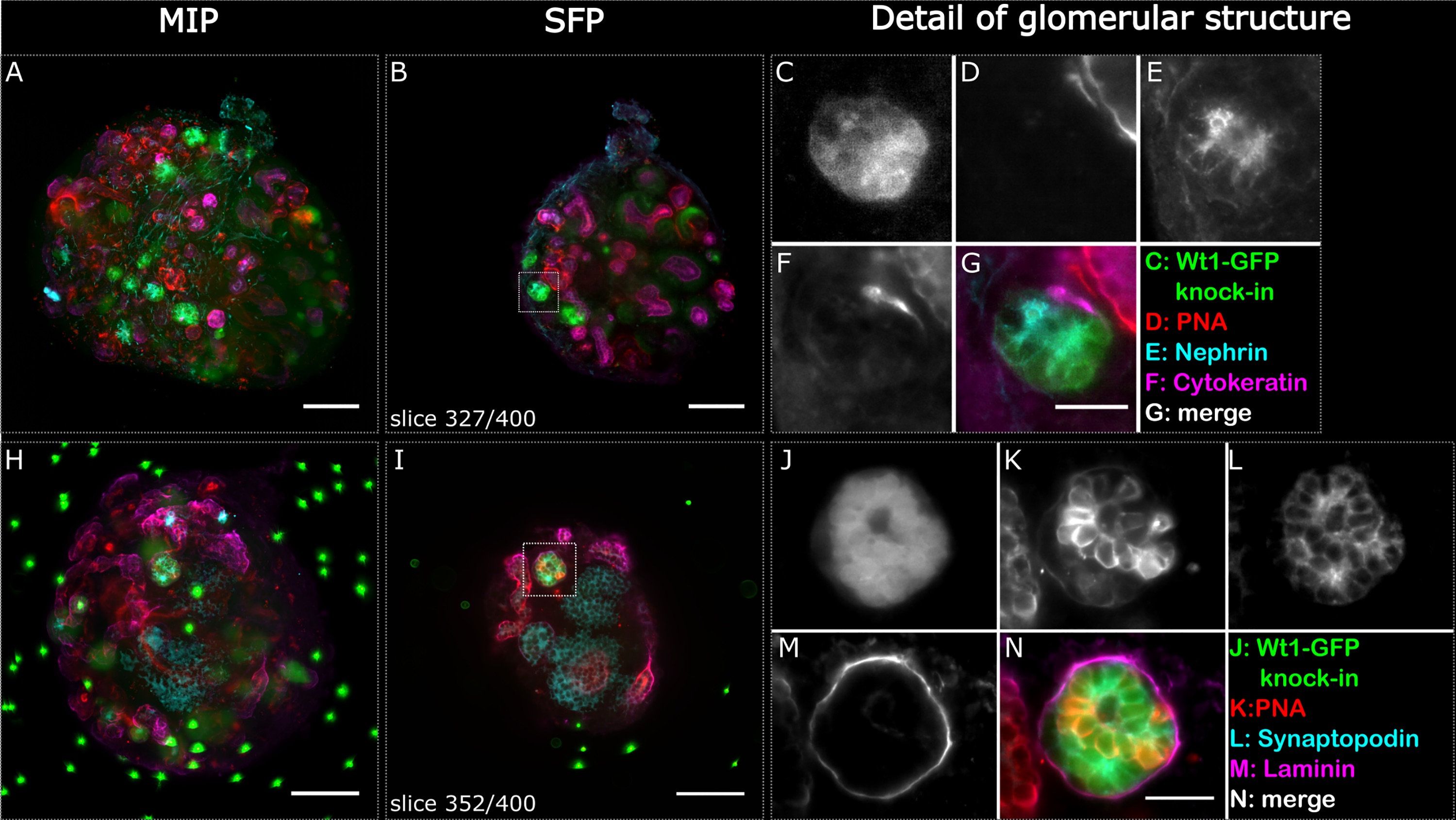
Renal Glomerular Organoids
Fig 2. Glomerular structures stained positively for podocyte markers indicating phenotypic maturity in re-aggregated embryonic renal organoids after 6 days of culture. Two whole organoids (A-G and H-N) were imaged in the light sheet microscope and are depicted as maximum intensity projections (A, H) and respective selected single focal planes (B, I). Glomerular structures are highlighted with a dashed square in the single focal plane, and detailed images are shown in C-G and J-N respectively. The Wt1-GFP structure in C also stained positive for nephrin (E), and the Wt1-GFP structure in J stained positive for synaptopodin (L). Cytokeratin highlights the various ureteric bud foci, and PNA and laminin the basement membranes of the ureteric bud and nephric tubules. The sample shown in H-N contains fluorescent beads as reference points for multi view reconstruction of the dataset. The organoids were not cleared. Scale bars in A, B, H and I: 100 μm, G: 10 μm, N: 25μm. https://doi.org/10.1371/journal.pone.0199918.g002
File history
Click on a date/time to view the file as it appeared at that time.
| Date/Time | Thumbnail | Dimensions | User | Comment | |
|---|---|---|---|---|---|
| current | 05:51, 30 July 2019 | | 3,000 × 1,689 (700 KB) | Z8600021 (talk | contribs) | ==Renal Glomerular Organoids== Fig 2. Glomerular structures stained positively for podocyte markers indicating phenotypic maturity in re-aggregated embryonic renal organoids after 6 days of culture. Two whole organoids (A-G and H-N) were imaged in the... |
You cannot overwrite this file.
File usage
The following page uses this file: