File:Organoids - renal glomerulus.jpg
Original file (3,000 × 1,689 pixels, file size: 700 KB, MIME type: image/jpeg)
Renal Glomerular Organoids
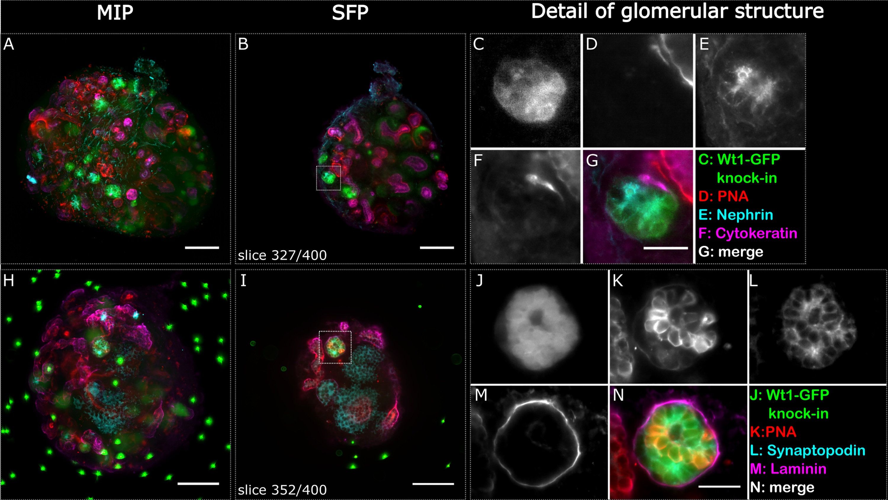
Fig 2. Glomerular structures stained positively for podocyte markers indicating phenotypic maturity in re-aggregated embryonic renal organoids after 6 days of culture. Two whole organoids (A-G and H-N) were imaged in the light sheet microscope and are depicted as maximum intensity projections (A, H) and respective selected single focal planes (B, I). Glomerular structures are highlighted with a dashed square in the single focal plane, and detailed images are shown in C-G and J-N respectively. The Wt1-GFP structure in C also stained positive for nephrin (E), and the Wt1-GFP structure in J stained positive for synaptopodin (L). Cytokeratin highlights the various ureteric bud foci, and PNA and laminin the basement membranes of the ureteric bud and nephric tubules. The sample shown in H-N contains fluorescent beads as reference points for multi view reconstruction of the dataset. The organoids were not cleared. Scale bars in A, B, H and I: 100 μm, G: 10 μm, N: 25μm.
Reference
Held M, Santeramo I, Wilm B, Murray P & Lévy R. (2018). Ex vivo live cell tracking in kidney organoids using light sheet fluorescence microscopy. PLoS ONE , 13, e0199918. PMID: 30048451 DOI.
Copyright
© 2018 Held et al. This is an open access article distributed under the terms of the Creative Commons Attribution License, which permits unrestricted use, distribution, and reproduction in any medium, provided the original author and source are credited.
https://doi.org/10.1371/journal.pone.0199918.g002
Cite this page: Hill, M.A. (2024, April 30) Embryology Organoids - renal glomerulus.jpg. Retrieved from https://embryology.med.unsw.edu.au/embryology/index.php/File:Organoids_-_renal_glomerulus.jpg
- © Dr Mark Hill 2024, UNSW Embryology ISBN: 978 0 7334 2609 4 - UNSW CRICOS Provider Code No. 00098G
File history
Click on a date/time to view the file as it appeared at that time.
| Date/Time | Thumbnail | Dimensions | User | Comment | |
|---|---|---|---|---|---|
| current | 05:51, 30 July 2019 | | 3,000 × 1,689 (700 KB) | Z8600021 (talk | contribs) | ==Renal Glomerular Organoids== Fig 2. Glomerular structures stained positively for podocyte markers indicating phenotypic maturity in re-aggregated embryonic renal organoids after 6 days of culture. Two whole organoids (A-G and H-N) were imaged in the... |
You cannot overwrite this file.
File usage
The following page uses this file: