File:Odgers1939-fig04.jpg
From Embryology
Size of this preview: 800 × 586 pixels. Other resolution: 1,000 × 732 pixels.
Original file (1,000 × 732 pixels, file size: 250 KB, MIME type: image/jpeg)
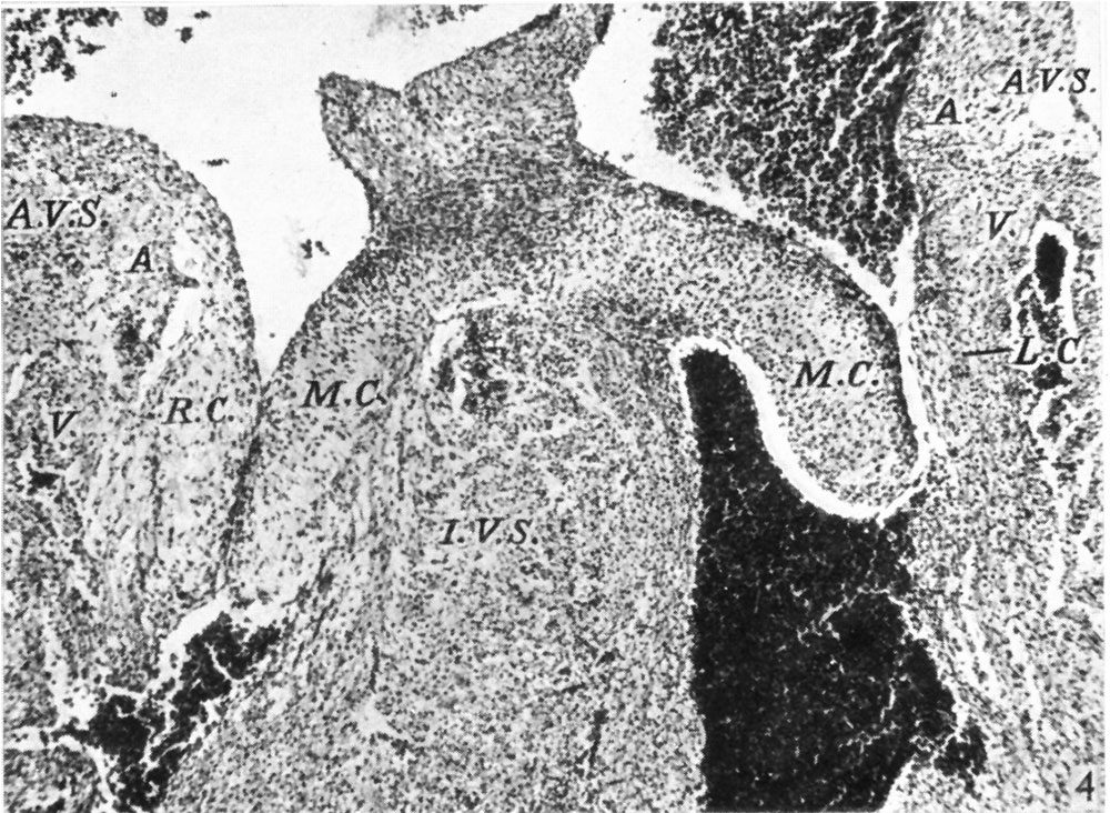
Fig. 4. A section through the heart in a 23 mm. embryo
( x 60). It shows the cushions of the central cusps, M .0., capping the muscular interventricular septum, I .V.S., and those of the lateral cusps, 12.0’. and L0. Note the angulation of the right A.-V. sulcus, A.V.S. A. auricular, V. ventricular muscle.
File history
Click on a date/time to view the file as it appeared at that time.
| Date/Time | Thumbnail | Dimensions | User | Comment | |
|---|---|---|---|---|---|
| current | 15:00, 15 November 2015 | | 1,000 × 732 (250 KB) | Z8600021 (talk | contribs) |
You cannot overwrite this file.
File usage
The following 3 pages use this file: