File:Odgers1938 fig11.jpg: Difference between revisions
No edit summary |
mNo edit summary |
||
| (One intermediate revision by the same user not shown) | |||
| Line 1: | Line 1: | ||
==Plate II== | |||
Abbreviations | |||
* R.B.R. right bulbar ridge. | |||
* L.B.R. left bulbar ridge. | |||
* R.S.T. right superior tubercle of the A.-V. cushions. | |||
* R.I.T. right inferior tubercle of the A.-V. cushions. | |||
* I.V.S. interventricular septum. | |||
* A.V.C. auriculoventricular cushions. | |||
* B.A.C. bulbo-auricular channel. | |||
Fig. 11. This is a section through the heart of a 17 mm. embryo ( x 80) five sections below the | |||
closing foramen. It shows the A.-V. cushion, A.V.0., separated, as in the last figure, by a | |||
notch on the right side from the cushion tissue, X, which has proliferated from it to join the | |||
interventricular septum, I. 17.18’. | |||
{{Odgers1938 figures}} | |||
Latest revision as of 20:01, 22 April 2016
Plate II
Abbreviations
- R.B.R. right bulbar ridge.
- L.B.R. left bulbar ridge.
- R.S.T. right superior tubercle of the A.-V. cushions.
- R.I.T. right inferior tubercle of the A.-V. cushions.
- I.V.S. interventricular septum.
- A.V.C. auriculoventricular cushions.
- B.A.C. bulbo-auricular channel.
Fig. 11. This is a section through the heart of a 17 mm. embryo ( x 80) five sections below the
closing foramen. It shows the A.-V. cushion, A.V.0., separated, as in the last figure, by a
notch on the right side from the cushion tissue, X, which has proliferated from it to join the
interventricular septum, I. 17.18’.
| Historic Disclaimer - information about historic embryology pages |
|---|
 |
Reference
Odgers PN. The development of the pars membranacea septi in the human heart. (1938) J Anat. 72(2): 247-59. https://www.ncbi.nlm.nih.gov/pubmed/17104688 PMID 17104688]
Cite this page: Hill, M.A. (2024, May 19) Embryology Odgers1938 fig11.jpg. Retrieved from https://embryology.med.unsw.edu.au/embryology/index.php/File:Odgers1938_fig11.jpg
- © Dr Mark Hill 2024, UNSW Embryology ISBN: 978 0 7334 2609 4 - UNSW CRICOS Provider Code No. 00098G
File history
Click on a date/time to view the file as it appeared at that time.
| Date/Time | Thumbnail | Dimensions | User | Comment | |
|---|---|---|---|---|---|
| current | 11:33, 25 January 2016 | 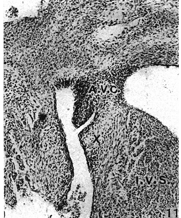 | 587 × 708 (166 KB) | Z8600021 (talk | contribs) |
You cannot overwrite this file.
File usage
The following 2 pages use this file: