File:Odgers1935 textfig04.jpg

Original file (900 × 698 pixels, file size: 139 KB, MIME type: image/jpeg)
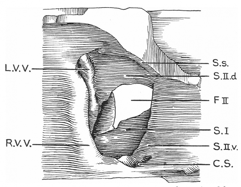
Text-fig. 4. Drawing of a model of the auricles in a 28.5 mm embryo
Viewed from the right side, to show the venous valves and the septal formations. SJ. septum primum; S.s. septum spurium; F.II. foramen secundum; R.V.V. right venous valve; L.V.V. left venous valve; C.S. coronary sinus; S.II.d. septum secundum, dorsal segment; S.Il .12. septum secundum, ventral segment.
Reference
Odgers PNB. The formation of the venous valves, the foramen secundum and the septum secundum in the human heart. (1935) J. Anat., 69: 412-422. PMID 17104548
Cite this page: Hill, M.A. (2024, April 27) Embryology Odgers1935 textfig04.jpg. Retrieved from https://embryology.med.unsw.edu.au/embryology/index.php/File:Odgers1935_textfig04.jpg
- © Dr Mark Hill 2024, UNSW Embryology ISBN: 978 0 7334 2609 4 - UNSW CRICOS Provider Code No. 00098G
File history
Click on a date/time to view the file as it appeared at that time.
| Date/Time | Thumbnail | Dimensions | User | Comment | |
|---|---|---|---|---|---|
| current | 14:44, 5 March 2017 | | 900 × 698 (139 KB) | Z8600021 (talk | contribs) | |
| 14:44, 5 March 2017 | 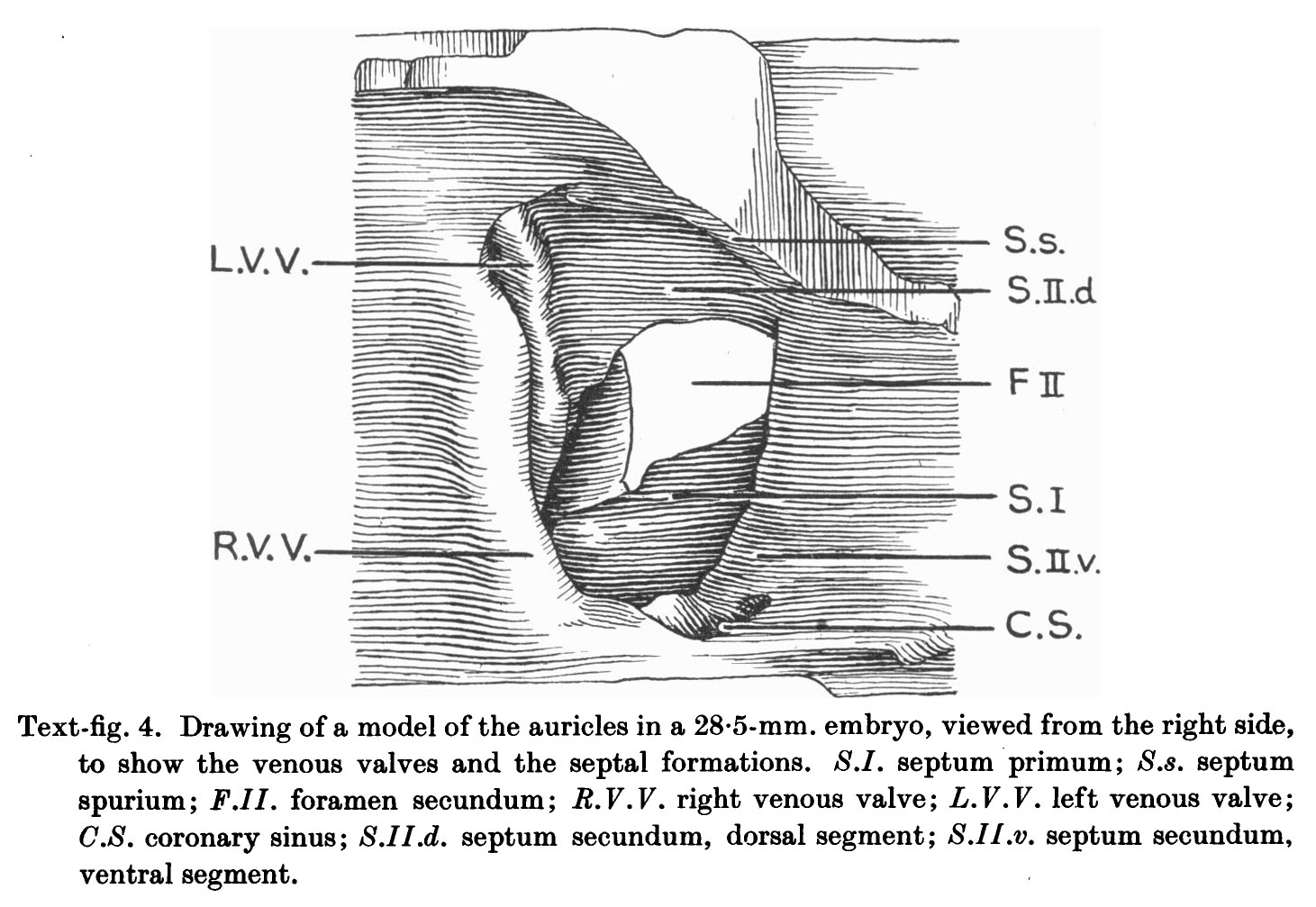 | 1,461 × 1,007 (232 KB) | Z8600021 (talk | contribs) | ===Reference=== {{Ref-Odgers1935}} {{Footer}} |
You cannot overwrite this file.
File usage
The following page uses this file: