File:O'Rahilly1957 plate01.jpg

Original file (2,105 × 2,990 pixels, file size: 1.22 MB, MIME type: image/jpeg)
Plate 1
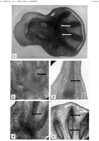
Fig. 1. Frontal section of hand of embryo, Carnegie no. 8789, 11.7 mm., horizon xvii. X56. Senior's stage 1. lilastemal condensations of digits 2 to 5. Radius and ulna are chondrifying. Carpus is blastemal, except very early chondrification in metacarpals 3 and 4, barely suggested in this figure by lighter-staining areas (arrows).
Fig. 2. Frontal section of left hand of embryo, Wayne University no. 15, not included in text because the embryo has not been assigned to a Streeter horizon. x75. Senior’s stage 5. Metacarpals 3 to 5 in the upper part of the figure. Hamate indicated by arrow; capitate and trapezoid to its left; triquetrum just below at its right. Radius and ulna in lower part of figure.
Fig. 3. Sagittal section of right foot of embryo, Wayne University no. 15. x75. Senior's stage 1 (early). Early chondrification in metatarsal 4 (arrow). A blastemal condensation for the I cuboid is visible as a more darkly stained region just below the metatarsal.
Fig. 4. Horizontal section of left foot of embryo, Wayne University no. 15. x75. Senior's stage 1 (early). Condensations of metatarsals 2 to 4 in upper part of figure (arrow indicates metatarsal 3). Lighter staining, as compared with tarsus (more darkly stained region just below metatarsals), suggests beginning chondrilication.
Fig. 5. Horizontal section of left foot of embryo, Wayne University no. 17-2, not included in text because the embryo has not been assigned to a Streeter horizon. x75. Senior's stage 7. Chondrification in metatarsals 2 to 4. Section includes proximal phalanges 3 and 4. Arrows indicate metatarsal 3 and proximal phalanx 3. Part of marginal vein is present above.
| Historic Disclaimer - information about historic embryology pages |
|---|
| Pages where the terms "Historic" (textbooks, papers, people, recommendations) appear on this site, and sections within pages where this disclaimer appears, indicate that the content and scientific understanding are specific to the time of publication. This means that while some scientific descriptions are still accurate, the terminology and interpretation of the developmental mechanisms reflect the understanding at the time of original publication and those of the preceding periods, these terms, interpretations and recommendations may not reflect our current scientific understanding. (More? Embryology History | Historic Embryology Papers) |
Reference
O'Rahilly R. Gray DI. and Gardner E. Chondrification in the hands and feet of staged human embryos. (1957) Carnegie Instn. Wash. Publ. 611, Contrib. Embryol., 36:
Cite this page: Hill, M.A. (2024, April 27) Embryology O'Rahilly1957 plate01.jpg. Retrieved from https://embryology.med.unsw.edu.au/embryology/index.php/File:O%27Rahilly1957_plate01.jpg
- © Dr Mark Hill 2024, UNSW Embryology ISBN: 978 0 7334 2609 4 - UNSW CRICOS Provider Code No. 00098G
File history
Click on a date/time to view the file as it appeared at that time.
| Date/Time | Thumbnail | Dimensions | User | Comment | |
|---|---|---|---|---|---|
| current | 17:01, 5 June 2016 |  | 2,105 × 2,990 (1.22 MB) | Z8600021 (talk | contribs) | |
| 16:53, 5 June 2016 |  | 2,105 × 2,990 (1.66 MB) | Z8600021 (talk | contribs) | ==Plate 1== {{Historic Disclaimer}} ===Reference=== {{Ref-O'Rahilly1957}} {{Footerr}} |
You cannot overwrite this file.
File usage
The following page uses this file:
