File:Normal control muscle (a) vs. Duchennes muscular dystrophy muscle (b).jpg: Difference between revisions
No edit summary |
|||
| (One intermediate revision by the same user not shown) | |||
| Line 5: | Line 5: | ||
Copyright notice: | Copyright notice: | ||
This is an open access article distributed under the Creative Commons Attribution License, which permits unrestricted use, distribution, and reproduction in any medium, provided the original work is properly cited. | This is an open access article distributed under the Creative Commons Attribution License, which permits unrestricted use, distribution, and reproduction in any medium, provided the original work is properly cited. | ||
===Assessment=== | |||
+ Good to show actual immunofluorescence of protein localisation. | |||
- No citation included image information as required by file criteria, this is a major error. | |||
- Clumsy file naming is a minor eror. | |||
{{Template:2011 Student Image}} | {{Template:2011 Student Image}} | ||
_vs._Duchennes_muscular_dystrophy_muscle_(b).jpg&oldid=77233)
_vs._Duchennes_muscular_dystrophy_muscle_(b).jpg&action=edit&oldid=77233)
_vs._Duchennes_muscular_dystrophy_muscle_(b).jpg&diff=prev&oldid=77233)
_vs._Duchennes_muscular_dystrophy_muscle_(b).jpg&oldid=79436)
_vs._Duchennes_muscular_dystrophy_muscle_(b).jpg&action=edit)
Latest revision as of 16:11, 24 October 2011
Immunofluorescence with anti-AQP4 antibody of normal control muscle (a) and Duchenne muscular dystrophy muscle (DMD) (b), and that with anti-spectrin antibody of serial muscle section of DMD (c). Positive immunoreactivity with anti-AQP4 antibody is seen in apparently all myofibers of normal control muscles (a); while it is noted in mosaic pattern in DMD muscle (b). DMD muscle contains less numerous myofibers with positive immunoreactivity of anti-AQP4 antibody (b) than myofibers with that of anti-spectrin antibody (c). Scattered anti-AQP4 immunonegative fibers (asterisks in (b)) are noted in DMD muscle. Scale bar in (a)–(c) = 50μm.
Normal control muscle (a) vs. Duchennes muscular dystrophy muscle (b).jpg
Copyright notice: This is an open access article distributed under the Creative Commons Attribution License, which permits unrestricted use, distribution, and reproduction in any medium, provided the original work is properly cited.
Assessment
+ Good to show actual immunofluorescence of protein localisation. - No citation included image information as required by file criteria, this is a major error. - Clumsy file naming is a minor eror.
- Note - This image was originally uploaded as part of a student project and may contain inaccuracies in either description or acknowledgements. Students have been advised in writing concerning the reuse of content and may accidentally have misunderstood the original terms of use. If image reuse on this non-commercial educational site infringes your existing copyright, please contact the site editor for immediate removal.
Cite this page: Hill, M.A. (2024, April 26) Embryology Normal control muscle (a) vs. Duchennes muscular dystrophy muscle (b).jpg. Retrieved from https://embryology.med.unsw.edu.au/embryology/index.php/File:Normal_control_muscle_(a)_vs._Duchennes_muscular_dystrophy_muscle_(b).jpg
_vs._Duchennes_muscular_dystrophy_muscle_(b).jpg)
_vs._Duchennes_muscular_dystrophy_muscle_(b).jpg)
- © Dr Mark Hill 2024, UNSW Embryology ISBN: 978 0 7334 2609 4 - UNSW CRICOS Provider Code No. 00098G
File history
Click on a date/time to view the file as it appeared at that time.
| Date/Time | Thumbnail | Dimensions | User | Comment | |
|---|---|---|---|---|---|
| current | 10:27, 22 September 2011 | 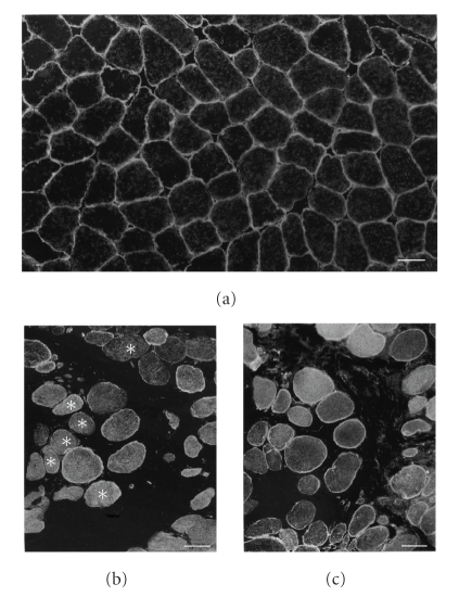 | 423 × 550 (131 KB) | Z3332629 (talk | contribs) | Immunofluorescence with anti-AQP4 antibody of normal control muscle (a) and Duchenne muscular dystrophy muscle (DMD) (b), and that with anti-spectrin antibody of serial muscle section of DMD (c). Positive immunoreactivity with anti-AQP4 antibody is seen i |
You cannot overwrite this file.
File usage
The following 2 pages use this file:
_vs._Duchennes_muscular_dystrophy_muscle_(b).jpg&oldid=79436)