File:Neutrophil EM01.jpg

Original file (999 × 1,000 pixels, file size: 402 KB, MIME type: image/jpeg)
Neutrophil Morphology Transmission electron microscopy (TEM)
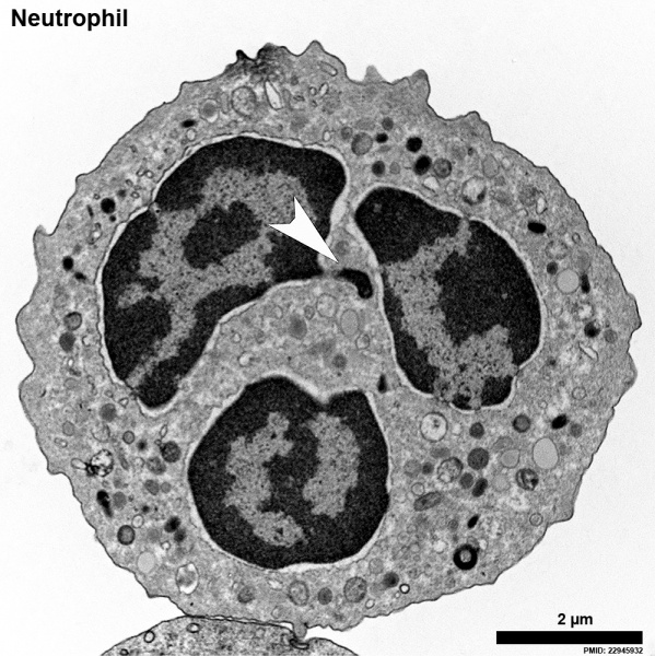
A naive human neutrophil contains various types of granules, clearly visible in the cytoplasm, as well as a lobulated nucleus. The highly condensed heterochromatin (dark) is neatly marginalized to the edge of the nucleus, only interrupted by euchromatic areas close to nuclear pores that mostly line the nuclear membrane. The brighter euchromatin is mostly in the center of the lobules.
White arrowhead - Barr body
This neutrophil comes from a female donor and one inactivated x chromosome can be found as an extranuclear stretch of heterochromatin. These structures are termed Barr bodies, and in neutrophils “drum sticks.”
Reference
<pubmed>22945932</pubmed>| J Cell Biol.
Copyright
Rockefeller University Press - Copyright Policy This article is distributed under the terms of an Attribution–Noncommercial–Share Alike–No Mirror Sites license for the first six months after the publication date (see http://www.jcb.org/misc/terms.shtml). After six months it is available under a Creative Commons License (Attribution–Noncommercial–Share Alike 4.0 Unported license, as described at https://creativecommons.org/licenses/by-nc-sa/4.0/ ). (More? Help:Copyright Tutorial)
Figure 1. altered in size, contrast and labelling.
File history
Click on a date/time to view the file as it appeared at that time.
| Date/Time | Thumbnail | Dimensions | User | Comment | |
|---|---|---|---|---|---|
| current | 09:22, 12 May 2015 |  | 999 × 1,000 (402 KB) | Z8600021 (talk | contribs) | ==Neutrophil Morphology Transmission electron microscopy (TEM)== A naive human neutrophil contains various types of granules, clearly visible in the cytoplasm, as well as a lobulated nucleus. The highly condensed heterochromatin (dark) is neatly margi... |
You cannot overwrite this file.
File usage
The following 3 pages use this file: