File:Nephron EM11.jpg: Difference between revisions
mNo edit summary |
|||
| Line 3: | Line 3: | ||
Electron Micrograph of the nephron glomerulus showing podocyte and glomerular capillary. | Electron Micrograph of the nephron glomerulus showing podocyte and glomerular capillary. | ||
This high magnification view shows podocytes with the interdigitated foot processes (pedicels) that are wrapped around the exterior of a glomerular capillary forming slit diaphragms (SDs). | This high magnification view shows podocytes with the interdigitated foot processes (pedicels) that are wrapped around the exterior of a single glomerular capillary. The interdigitated foot processes forming slit diaphragms (SDs). | ||
Scale Bar: 1 µm. | Scale Bar: 1 µm. | ||
'''Glomerular filtration barrier''' - formed by | '''Glomerular filtration barrier''' - formed by podocyte slit diaphragms and the fenestrated endothelium. | ||
:'''Links:''' [[:File:Nephron EM02.jpg|colour image version]] | {{renal histology}} | :'''Links:''' [[:File:Nephron EM02.jpg|colour image version]] | [[:File:Nephron EM1.jpg|smaller image]] | {{renal histology}} | ||
===Reference=== | ===Reference=== | ||
Revision as of 13:14, 24 July 2019
Glomerulus Podocyte Electron Micreograph
Electron Micrograph of the nephron glomerulus showing podocyte and glomerular capillary.
This high magnification view shows podocytes with the interdigitated foot processes (pedicels) that are wrapped around the exterior of a single glomerular capillary. The interdigitated foot processes forming slit diaphragms (SDs).
Scale Bar: 1 µm.
Glomerular filtration barrier - formed by podocyte slit diaphragms and the fenestrated endothelium.
- Links: colour image version | smaller image | renal histology
Reference
Scott RP & Quaggin SE. (2015). Review series: The cell biology of renal filtration. J. Cell Biol. , 209, 199-210. PMID: 25918223 DOI.
Copyright
Rockefeller University Press - Copyright Policy This article is distributed under the terms of an Attribution–Noncommercial–Share Alike–No Mirror Sites license for the first six months after the publication date (see http://www.jcb.org/misc/terms.shtml). After six months it is available under a Creative Commons License (Attribution–Noncommercial–Share Alike 4.0 Unported license, as described at https://creativecommons.org/licenses/by-nc-sa/4.0/ ). (More? Help:Copyright Tutorial)
Figure 2. Panel B cropped resized, relabelled and converted to black and white image. Text above modified from figure legend.
Cite this page: Hill, M.A. (2024, May 28) Embryology Nephron EM11.jpg. Retrieved from https://embryology.med.unsw.edu.au/embryology/index.php/File:Nephron_EM11.jpg
- © Dr Mark Hill 2024, UNSW Embryology ISBN: 978 0 7334 2609 4 - UNSW CRICOS Provider Code No. 00098G
File history
Click on a date/time to view the file as it appeared at that time.
| Date/Time | Thumbnail | Dimensions | User | Comment | |
|---|---|---|---|---|---|
| current | 13:12, 24 July 2019 | 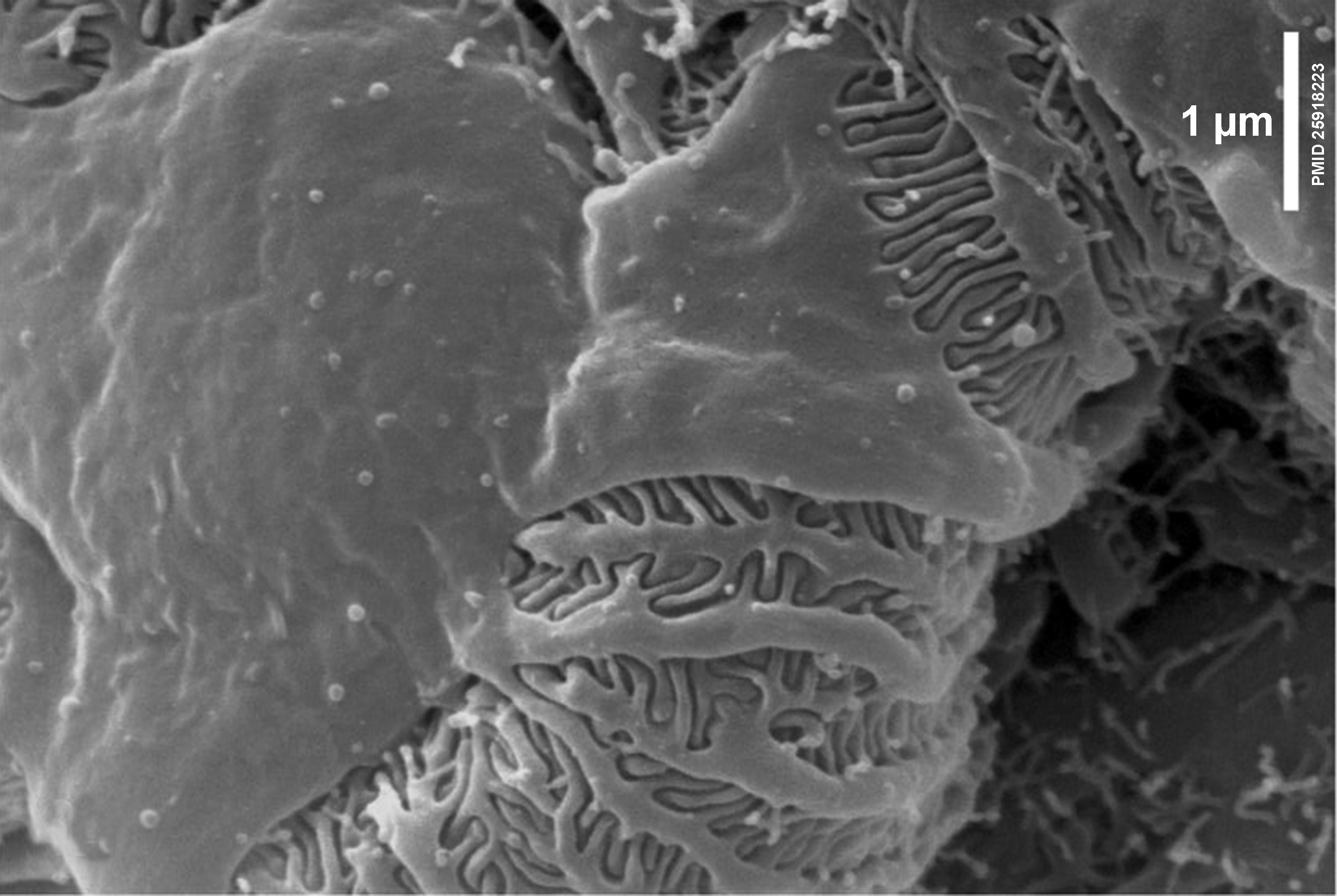 | 2,983 × 2,000 (398 KB) | Z8600021 (talk | contribs) | |
| 13:06, 24 July 2019 |  | 200 × 134 (9 KB) | Z8600021 (talk | contribs) |
You cannot overwrite this file.
File usage
There are no pages that use this file.