File:Neonatal human pulmonary neuroendocrine cell EM01.jpg
From Embryology

Size of this preview: 418 × 600 pixels. Other resolution: 836 × 1,200 pixels.
Original file (836 × 1,200 pixels, file size: 405 KB, MIME type: image/jpeg)
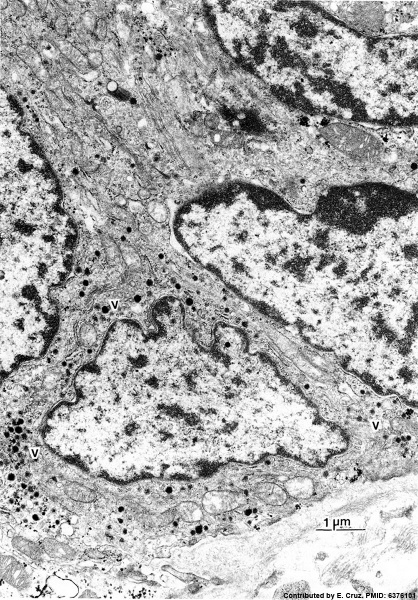
Human Pulmonary Neuroendocrine Cells (electron micrograph)
Portion of a neuroepithelial body in neonatal human airway epithelium showing three cells with dense-core vesicles (v).
TEM. x 14,000. Contributed by Dr. E. Cutz.
Reference
Copyright
Copyright: Reproduced with permission from Environmental Health Perspectives (EHP) is a publication of the U.S. Government. Publication of EHP lies in the public domain and is therefore without copyright.
Figure 9.
File history
Click on a date/time to view the file as it appeared at that time.
| Date/Time | Thumbnail | Dimensions | User | Comment | |
|---|---|---|---|---|---|
| current | 10:06, 13 October 2015 | | 836 × 1,200 (405 KB) | Z8600021 (talk | contribs) | |
| 10:04, 13 October 2015 |  | 836 × 1,200 (405 KB) | Z8600021 (talk | contribs) | ==Human Pulmonary Neuroendocrine Cells (electron micrograph)== Portion of a neuroepithelial body in neonatal human airway epithelium showing three cells with dense-core vesicles (v). TEM. x 14,000. Contributed by Dr. E. Cutz. ===Reference=== ====Co... |
You cannot overwrite this file.
File usage
The following 4 pages use this file: