File:Neonatal human pulmonary neuroendocrine cell EM01.jpg

Original file (836 × 1,200 pixels, file size: 405 KB, MIME type: image/jpeg)
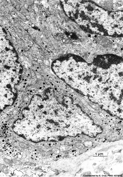
Human Pulmonary Neuroendocrine Cells (electron micrograph)
Portion of a neuroepithelial body in neonatal human airway epithelium showing three cells with dense-core vesicles (v).
TEM. x 14,000. Contributed by Dr. E. Cutz.
- Dense-core vesicles - occur in both nerve terminals and endocrine cells and can contain both neuropeptides with smaller neurotransmitters.
Reference
DiAugustine RP & Sonstegard KS. (1984). Neuroendocrinelike (small granule) epithelial cells of the lung. Environ. Health Perspect. , 55, 271-95. PMID: 6376101
Copyright
Copyright: Reproduced with permission from Environmental Health Perspectives (EHP) is a publication of the U.S. Government. Publication of EHP lies in the public domain and is therefore without copyright.
Figure 9. Original image adjusted in size, contrast and labelling.
Cite this page: Hill, M.A. (2024, April 27) Embryology Neonatal human pulmonary neuroendocrine cell EM01.jpg. Retrieved from https://embryology.med.unsw.edu.au/embryology/index.php/File:Neonatal_human_pulmonary_neuroendocrine_cell_EM01.jpg
- © Dr Mark Hill 2024, UNSW Embryology ISBN: 978 0 7334 2609 4 - UNSW CRICOS Provider Code No. 00098G
File history
Click on a date/time to view the file as it appeared at that time.
| Date/Time | Thumbnail | Dimensions | User | Comment | |
|---|---|---|---|---|---|
| current | 10:06, 13 October 2015 | | 836 × 1,200 (405 KB) | Z8600021 (talk | contribs) | |
| 10:04, 13 October 2015 |  | 836 × 1,200 (405 KB) | Z8600021 (talk | contribs) | ==Human Pulmonary Neuroendocrine Cells (electron micrograph)== Portion of a neuroepithelial body in neonatal human airway epithelium showing three cells with dense-core vesicles (v). TEM. x 14,000. Contributed by Dr. E. Cutz. ===Reference=== ====Co... |
You cannot overwrite this file.
File usage
The following 4 pages use this file: