File:Nelsen1953 fig006.jpg: Difference between revisions
From Embryology
(Z8600021 uploaded a new version of File:Nelsen1953 fig006.jpg) |
mNo edit summary |
||
| Line 1: | Line 1: | ||
==Fig. 6. == | ==Fig. 6. Schematic drawing of the testis and its relationship within the scrotum== | ||
On the right side of the drawing the muscle and connective-tissue layers surrounding the inguinal bursa and testis are shown; on the left side may be seen the loops of the cremaster muscle surrounding the tunica vaginalis communis. | |||
Revision as of 14:49, 26 October 2016
Fig. 6. Schematic drawing of the testis and its relationship within the scrotum
On the right side of the drawing the muscle and connective-tissue layers surrounding the inguinal bursa and testis are shown; on the left side may be seen the loops of the cremaster muscle surrounding the tunica vaginalis communis.
Reference
Nelsen OE. Comparative embryology of the vertebrates (1953) Mcgraw-Hill Book Company, New York.
Cite this page: Hill, M.A. (2024, May 18) Embryology Nelsen1953 fig006.jpg. Retrieved from https://embryology.med.unsw.edu.au/embryology/index.php/File:Nelsen1953_fig006.jpg
- © Dr Mark Hill 2024, UNSW Embryology ISBN: 978 0 7334 2609 4 - UNSW CRICOS Provider Code No. 00098G
File history
Click on a date/time to view the file as it appeared at that time.
| Date/Time | Thumbnail | Dimensions | User | Comment | |
|---|---|---|---|---|---|
| current | 14:28, 26 October 2016 | 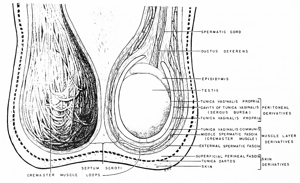 | 1,200 × 732 (224 KB) | Z8600021 (talk | contribs) | |
| 14:27, 26 October 2016 |  | 1,200 × 732 (211 KB) | Z8600021 (talk | contribs) | ||
| 14:27, 26 October 2016 |  | 2,011 × 1,464 (516 KB) | Z8600021 (talk | contribs) |
You cannot overwrite this file.
File usage
The following 2 pages use this file: