File:Muscle-reorganization microtububules and centrosome protein.jpg

Original file (1,000 × 1,125 pixels, file size: 140 KB, MIME type: image/jpeg)
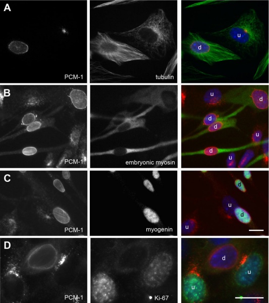
Reorganization of microtububules and centrosome protein in cells expressing myogenic differentiation markers.
Culture of mouse myoblasts containing undifferentiated (u) cells, and cells that started to differentiate (d).
The centrosome protein PCM-1 is stained in red, DNA is stained in blue. In green is marked (A) tubulin, (B) the differentiation marker 'embryonic myosin', (C) the differentiation marker myogenin, (D) the proliferation marker Ki-67.
Bars, 10 μm. Identical magnifications in A-C.
Original file name: 1471-2121-10-28-2-l.jpg http://www.biomedcentral.com/1471-2121/10/28/figure/F2
Original Figure cropped and resolution changed.
Centrosome proteins form an insoluble perinuclear matrix during muscle cell differentiation. Srsen V, Fant X, Heald R, Rabouille C, Merdes A. BMC Cell Biol. 2009 Apr 21;10:28. PMID: 19383121 | BMC Cell Biol.
© 2009 Srsen et al; licensee BioMed Central Ltd.
This is an Open Access article distributed under the terms of the Creative Commons Attribution License (http://creativecommons.org/licenses/by/2.0), which permits unrestricted use, distribution, and reproduction in any medium, provided the original work is properly cited.
File history
Click on a date/time to view the file as it appeared at that time.
| Date/Time | Thumbnail | Dimensions | User | Comment | |
|---|---|---|---|---|---|
| current | 12:20, 1 May 2010 | | 1,000 × 1,125 (140 KB) | S8600021 (talk | contribs) | Reorganization of microtububules and centrosome protein in cells expressing myogenic differentiation markers. Culture of mouse myoblasts containing undifferentiated (u) cells, and cells that started to differentiate (d). The centrosome protein PCM-1 is |
You cannot overwrite this file.
File usage
There are no pages that use this file.