File:Mouse yolk sac 01.jpg

Original file (1,002 × 669 pixels, file size: 115 KB, MIME type: image/jpeg)
Mouse Yolk Sac Development
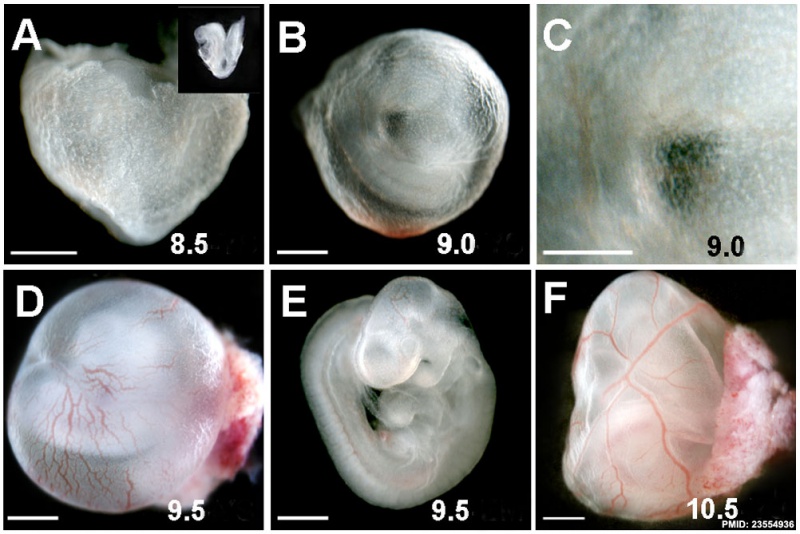
Bright field images of wild-type WT embryos alone or embryos within their yolk sacs at the indicated stages.
Prominent large blood vessels are easily detected in 10.5 dpc WT yolk sacs.
Reference
<pubmed>23554936</pubmed>| PMC3598950 | PLoS One.
Copyright
Copyright: © 2013 Rhee et al. This is an open-access article distributed under the terms of the Creative Commons Attribution License, which permits unrestricted use, distribution, and reproduction in any medium, provided the original author and source are credited.
Fig. 1. http://www.ncbi.nlm.nih.gov/pmc/articles/PMC3598950/figure/pone-0058828-g001 Wild-type embryo and yolk sac cropped from full figure, resized and relabelled)
File history
Click on a date/time to view the file as it appeared at that time.
| Date/Time | Thumbnail | Dimensions | User | Comment | |
|---|---|---|---|---|---|
| current | 16:45, 24 May 2013 |  | 1,002 × 669 (115 KB) | Z8600021 (talk | contribs) | PLoS One. 2013;8(3):e58828. doi: 10.1371/journal.pone.0058828. Epub 2013 Mar 15. Visceral endoderm expression of Yin-Yang1 (YY1) is required for VEGFA maintenance and yolk sac development. Rhee S, Guerrero-Zayas MI, Wallingford MC, Ortiz-Pineda P, Mage... |
You cannot overwrite this file.
File usage
There are no pages that use this file.