File:Mouse spermatozoa mito movie icon.jpg
Mouse_spermatozoa_mito_movie_icon.jpg (495 × 495 pixels, file size: 43 KB, MIME type: image/jpeg)
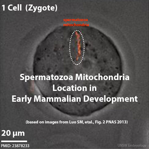
Spermatozoa Mitochondria in Early Mouse Embryos
Image has been labeled for movie icon display.
Live cell fluorescence imaging of sperm mitochondria (red) in early embryos. Sperm mitochondria disaggregated and became restricted to only one blastomere during one-cell to four-cell stages. Sperm mitochondria distributed in several cells after the eight-cell stage and could be detected until morula stages.
Scale bars: 20 µm.
- Links: Mitochondria | Spermatozoa Development | Fertilization
Reference
<pubmed>23878233</pubmed>| Proc Natl Acad Sci U S A.
Copyright
Proceedings National Academy of Sciences (PNAS) Liberalization of PNAS copyright policy: Noncommercial use freely allowed Note original Author should be contacted for permission to reuse for Educational purposes. See also PNAS Author Rights and Permission FAQs
- Cozzarelli NR, Fulton KR, Sullenberger DM. Liberalization of PNAS copyright policy: noncommercial use freely allowed. Proc Natl Acad Sci U S A. 2004 Aug 24;101(34):12399. PMID15314225 "Our guiding principle is that, while PNAS retains copyright, anyone can make noncommercial use of work in PNAS without asking our permission, provided that the original source is cited."
Fig. 2. http://www.pnas.org/content/110/32/13038/F2.expansion.html Original image resized and relabeled.
File history
Click on a date/time to view the file as it appeared at that time.
| Date/Time | Thumbnail | Dimensions | User | Comment | |
|---|---|---|---|---|---|
| current | 13:46, 8 April 2014 | 495 × 495 (43 KB) | Z8600021 (talk | contribs) | ==Spermatozoa Mitochondria in Early Mouse Embryos== Image has been labeled for movie icon display. Live cell fluorescence imaging of sperm mitochondria (red) in early embryos. Sperm mitochondria disaggregated and became restricted to only one blasto... |
You cannot overwrite this file.
File usage
The following 32 pages use this file:
- 2016Lecture-Gametogenesis-Fertilization-Movie
- ANAT2341 Lab 1 - Fertilization
- Animal Development
- Animations
- BGDA Practical 3 - Fertilization
- Book - The maturation of the egg of the mouse (1911)
- Lecture - Fertilization
- Mitochondria
- Models of Human Development
- Mouse Development
- Mouse E13 microCT Movie
- Mouse Estrous Cycle
- Mouse Heart
- Mouse Stages
- Mouse Timeline
- Mouse Timeline Detailed
- Movies
- Paper - Development of the Mouse Gonads 1
- Paper - Development of the Mouse Gonads 2
- Paper - Development of the Mouse Gonads 3
- Paper - Development of the Mouse Gonads 4
- Paper - Oogenesis in the white mouse (1917)
- Paper - The development of the ear-bones in the mouse
- Paper - The involution of the transitory cortex of the mouse suprarenal (1933)
- Paper - The oestrous cycle in the mouse (1922)
- Paper - The prenatal growth of the mouse
- File:Mouse spermatozoa mitochondria 01.jpg
- Template:Mitochondria movie 1
- Template:Mouse links
- Category:Mouse
- Category:Mouse E12.5
- Category:Mouse E14.5