File:Mouse pancreas development.jpg
Original file (600 × 939 pixels, file size: 261 KB, MIME type: image/jpeg)
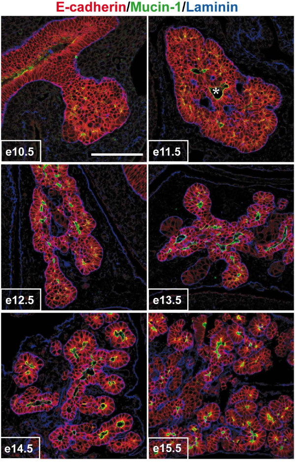
Mouse Pancreas Development
Remodeling of the pancreatic epithelial cell mass into polarized monolayers.
- Pancreatic sections from e10.5 to e15.5 embryos were examined by immunofluorescence using antibodies directed against E-cadherin, mucin-1 and laminin.
- Pancreatic development starts by the formation of a mass of non-polarized epithelial cells (e10.5-e11.5).
- This mass is then remodeled (e12.5-e13.5) and finally every epithelial cell become polarized, with its basal pole contacting laminin and its apical pole facing a lumen (e14.5-e15.5).
- * indicates the central duct, in connection with the duodenum.
Scale bar, 100 μm.
Original file name: 1471-213X-9-66-1.jpg
Reference
<pubmed>20003423</pubmed>| BMC Dev Biol.
Hick et al. BMC Developmental Biology 2009 9:66 doi:10.1186/1471-213X-9-66
© 2009 Hick et al; licensee BioMed Central Ltd.
This is an Open Access article distributed under the terms of the Creative Commons Attribution License (http://creativecommons.org/licenses/by/2.0), which permits unrestricted use, distribution, and reproduction in any medium, provided the original work is properly cited.
File history
Click on a date/time to view the file as it appeared at that time.
| Date/Time | Thumbnail | Dimensions | User | Comment | |
|---|---|---|---|---|---|
| current | 19:07, 9 July 2011 | | 600 × 939 (261 KB) | S8600021 (talk | contribs) | ==Mouse Pancreas Development== Remodeling of the pancreatic epithelial cell mass into polarized monolayers. Pancreatic sections from e10.5 to e15.5 embryos were examined by immunofluorescence using antibodies directed against E-cadherin, mucin-1 and la |
You cannot overwrite this file.
File usage
The following page uses this file: