File:Mouse organ of corti NeuroD1.jpg

Original file (1,779 × 2,383 pixels, file size: 1.11 MB, MIME type: image/jpeg)
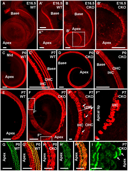
Neurod1 is necessary for development of an orderly patterned organ of Corti
Myo VIIa immunocytochemistry shows upregulation of Myo VIIa in the wild-type starts around E16.5 from the mid-base and later progresses toward both base and apex with a regular organization of one row of inner and three rows of outer hair cells throughout the cochlea (A, A’, C, C’, E). In contrast, Myo VIIa is already expressed throughout the cochlea with disorganization of hair cells in the apical half (B, B’, D) in E16.5 Neurod1 CKO mutant littermates. In later stages, the basal half of the CKO mutant shows normal orientation of the hair cells (D, D’) whereas the apex of Neurod1 CKO mice shows multiple rows of both IHCs and OHCs with reduction of Myo VIIa intensity in most outer hair cells (F, F’). In addition, clusters of higher intensity of Myo VIIa positive cells are found in between outer hair cells with equivalent staining intensity to inner hair cells (arrows in F’). The apical tip of the mutant cochlea shows a partially duplicated row of inner hair cells with complete absence of outer hair cells (F”). Using espin immunocytochemistry we confirm the disorganization of the apical half of the mutant cochlea where two rows of inner hair stereocilia and four to five rows of outer hair stereocilia are observed (H–H’) along with some unusually displaced strongly stained inner hair stereocilia (arrow in I) in between faintly labeled outer hair stereocilia (I). IHC, inner hair cells; OHC, outer hair cells. Bar indicates 100 µm except F”; 10 µm in F”.
Reference
Copyright
Copyright: © 2010 Jahan et al. This is an open-access article distributed under the terms of the Creative Commons Attribution License, which permits unrestricted use, distribution, and reproduction in any medium, provided the original author and source are credited.
Figure 5. https://doi.org/10.1371/journal.pone.0011661.g005
Above text modified from original figure legend.
Cite this page: Hill, M.A. (2024, May 8) Embryology Mouse organ of corti NeuroD1.jpg. Retrieved from https://embryology.med.unsw.edu.au/embryology/index.php/File:Mouse_organ_of_corti_NeuroD1.jpg
- © Dr Mark Hill 2024, UNSW Embryology ISBN: 978 0 7334 2609 4 - UNSW CRICOS Provider Code No. 00098G
File history
Click on a date/time to view the file as it appeared at that time.
| Date/Time | Thumbnail | Dimensions | User | Comment | |
|---|---|---|---|---|---|
| current | 18:24, 30 July 2018 |  | 1,779 × 2,383 (1.11 MB) | Z8600021 (talk | contribs) | Figure 5. Neurod1 is necessary for development of an orderly patterned organ of Corti. Myo VIIa immunocytochemistry shows upregulation of Myo VIIa in the wild-type starts around E16.5 from the mid-base and later progresses toward both base and apex wit... |
You cannot overwrite this file.
File usage
The following page uses this file: