File:Mouse oocyte cortical granules 02.jpg
Original file (1,006 × 1,000 pixels, file size: 177 KB, MIME type: image/jpeg)
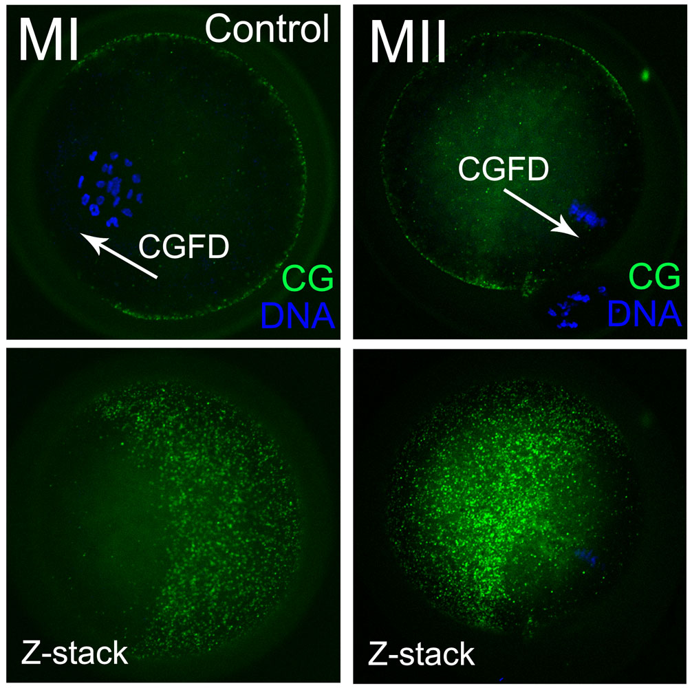
Mouse Oocyte Cortical Granules
The cortical granules were absent in the cortex close to where the chromosomes were located during the MI and MII stages in the control group. Note MI and MII images are of different oocytes.
- Z-stack showed the presence of different scanned layers.
- An arrowhead shows the cortical granule-free domain.
- Green - cortical granules.
- Blue - chromatin.
- Bar = 20 µm.
Reference
<pubmed>21494665</pubmed>| PLoS One.
Sun S-C, Wang Z-B, Xu Y-N, Lee S-E, Cui X-S, et al. (2011) Arp2/3 Complex Regulates Asymmetric Division and Cytokinesis in Mouse Oocytes. PLoS ONE 6(4): e18392. doi:10.1371/journal.pone.0018392
Copyright: © 2011 Sun et al. This is an open-access article distributed under the terms of the Creative Commons Attribution License, which permits unrestricted use, distribution, and reproduction in any medium, provided the original author and source are credited.
original file name Journal.pone.0018392.g006.jpg Figure 6. Effects of CK666 treatment and RNAi on cortical granule-free domain formation in mouse oocytes.
Control panels were cropped and resized from the full figure.
File history
Click on a date/time to view the file as it appeared at that time.
| Date/Time | Thumbnail | Dimensions | User | Comment | |
|---|---|---|---|---|---|
| current | 16:18, 6 May 2012 | | 1,006 × 1,000 (177 KB) | Z8600021 (talk | contribs) | ==Mouse Oocyte Cortical Granules== The cortical granules were absent in the cortex close to where the chromosomes were located during the MI and MII stages in the control group. Conversely, in the oocytes treated with CK666 and RNAi, the cortical granul |
You cannot overwrite this file.
File usage
The following page uses this file: