File:Mouse oocyte Egr3-01.jpg

Original file (600 × 670 pixels, file size: 152 KB, MIME type: image/jpeg)
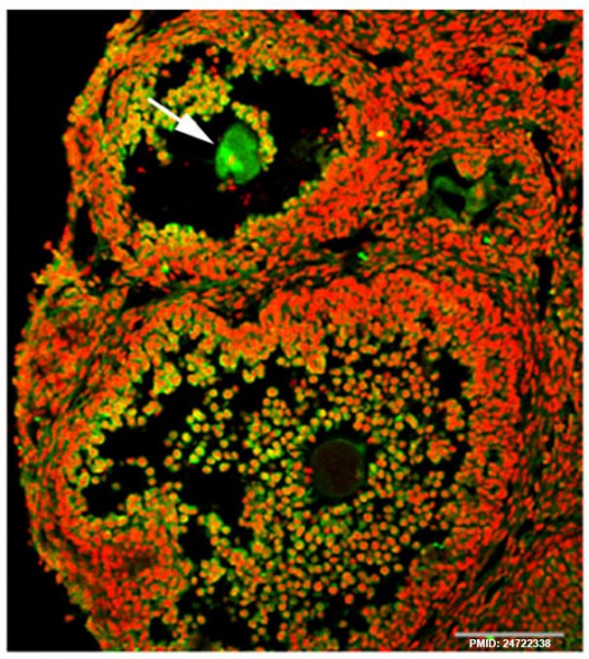
Mouse Oocyte Egr3 Expression
Egr3 localization in a mouse oocyte within a growing follicle. Meiotic spindles of maturing oocytes (Fig. 1A, arrow).
Immunofluorescence staining of Egr3 was performed on ovarian cryosection fixed in acetone. The rabbit polyclonal anti-Egr3 antibody (Santa Cruz) was used at 2 μg/ml. Primary antibody was probed with goat anti-rabbit IgG-Alexa Fluor 488 antibody. The DNA was counter-stained with TO-PRO-3-iodide.
Green, Egr3; red, DNA. White scale bar, 100 μm.
Reference
<pubmed>24722338</pubmed>| PLoS One.
Copyright
© 2014 Shin et al. This is an open-access article distributed under the terms of the Creative Commons Attribution License, which permits unrestricted use, distribution, and reproduction in any medium, provided the original author and source are credited.
Citation: Shin H, Kwon S, Song H, Lim HJ (2014) The Transcription Factor Egr3 Is a Putative Component of the Microtubule Organizing Center in Mouse Oocytes. PLoS ONE 9(4): e94708. doi:10.1371/journal.pone.0094708
Journal.pone.0094708.g001.jpg Fig 1 cropped, resized and relabeled.
File history
Click on a date/time to view the file as it appeared at that time.
| Date/Time | Thumbnail | Dimensions | User | Comment | |
|---|---|---|---|---|---|
| current | 22:38, 16 April 2014 | | 600 × 670 (152 KB) | Z8600021 (talk | contribs) | While investigating the expression of Egr transcription factors in the mouse ovary, we unexpectedly observed that Egr3 was localized to the meiotic spindles of maturing oocytes (Fig. 1A, arrow). Journal.pone.0094708.g001.jpg |
You cannot overwrite this file.
File usage
There are no pages that use this file.