File:Mouse mammary development 01.jpg

Original file (1,200 × 773 pixels, file size: 147 KB, MIME type: image/jpeg)
Embryonic Mouse Mammary Development
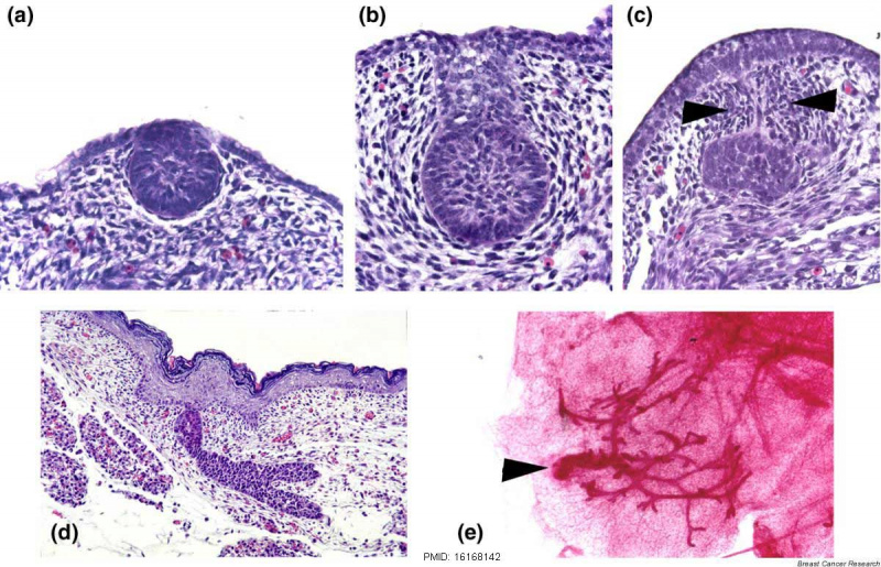
(a) Embryonic day E12.5 The epithelial cells have invaginated to form the initial bud, but the dense mammary mesenchyme has not yet formed.
(b) Embryonic day E14.5 - Female bud is fully formed. The epithelial cells are arrayed in a ball-on-stalk, or inverted bulb shape. The mesenchymal cells are arranged in four to five layers in a radial fashion around the epithelial cells.
(c) Embryonic day E14.5 Male bud under the influence of testosterone, the mesenchymal cells condense around the stalk of the bud (arrowheads), constricting it until the connection with the surface epidermis is severed. After this occurs mammary mesenchyme cells and many epithelial cells undergo apoptosis.
(d) Embryonic day E18.5 Mammary sprout epithelial bud has grown out from the mammary mesenchyme into the lower dermis, where it will enter the mammary fat pad and begin a period of active ductal branching morphogenesis.
(e) Mouse Day 2 - A whole mount of the initial primary duct system, the end- result of embryonic mammary morphogenesis. The arrowhead denotes the connection of the primary duct to the skin.
Reference
Hens JR & Wysolmerski JJ. (2005). Key stages of mammary gland development: molecular mechanisms involved in the formation of the embryonic mammary gland. Breast Cancer Res. , 7, 220-4. PMID: 16168142 DOI.
Copyright
The open access articles published in BioMed Central journals are made available under the Creative Commons Attribution (CC-BY) license, which means they are accessible online without any restrictions and can be re-used in any way, subject only to proper attribution (which, in an academic context, usually means citation).
See also - BMC Copyright Tutorial | BMC permissions
Fig. 1 - 13058_2005_1285_Fig1_HTML.jpg Text modified from original figure legend.
Key stages of mammary gland development: molecular mechanisms involved in the formation of the embryonic mammary gland.
Breast Cancer Res. 2005;7(5):220-4. Epub 2005 Aug 10.
Hens JR1, Wysolmerski JJ.
Abstract
The development of the embryonic mammary gland involves communication between the epidermis and mesenchyme and is coordinated temporally and spatially by various signaling pathways. Although many more genes are likely to control mammary gland development, functional roles have been identified for Wnt, fibroblast growth factor, and parathyroid hormone-related protein signaling. This review describes what is known about the molecular mechanisms that regulate embryonic mammary gland development.
PMID: 16168142 PMCID: PMC1242158 DOI: 10.1186/bcr1306
Cite this page: Hill, M.A. (2024, April 27) Embryology Mouse mammary development 01.jpg. Retrieved from https://embryology.med.unsw.edu.au/embryology/index.php/File:Mouse_mammary_development_01.jpg
- © Dr Mark Hill 2024, UNSW Embryology ISBN: 978 0 7334 2609 4 - UNSW CRICOS Provider Code No. 00098G
File history
Click on a date/time to view the file as it appeared at that time.
| Date/Time | Thumbnail | Dimensions | User | Comment | |
|---|---|---|---|---|---|
| current | 08:39, 20 March 2018 |  | 1,200 × 773 (147 KB) | Z8600021 (talk | contribs) | Embryonic mammary development. (a) Embryonic day (E)12.5. The epithelial cells have invaginated to form the initial bud, but the dense mammary mesenchyme has not yet formed. (b) Female bud at E14.5. The bud is fully formed. The epithelial cells are arr... |
You cannot overwrite this file.
File usage
There are no pages that use this file.