File:Mouse lung development 02.jpg: Difference between revisions
(==Histological analyses of Mouse Lungs at Various Embryonic Stages== Lung sections stained with H and E, and taken at various gestational stages as indicated. Images are at lower magnification (10X) The images are representative sections from wild-type) |
|||
| (One intermediate revision by the same user not shown) | |||
| Line 1: | Line 1: | ||
==Histological analyses of Mouse Lungs at Various Embryonic Stages== | ==Histological analyses of Mouse Lungs at Various Embryonic Stages== | ||
Lung sections stained with H and E, and taken at various gestational stages as indicated. | Lung sections stained with H and E, and taken at various gestational stages as indicated. The images are representative sections from wild-type (+/+) embryos, images are at lower magnification (10X) | ||
'''Labels:''' | |||
Labels: | |||
* de - distal epithelium | * de - distal epithelium | ||
* m - mesenchyme | * m - mesenchyme | ||
| Line 12: | Line 10: | ||
* b - bronchi | * b - bronchi | ||
:'''Links:''' [[:File:Mouse lung development 01.jpg|Full image]] | |||
Original file name: 1471-213X-4-1-3.jpg http://www.biomedcentral.com/1471-213X/4/1/figure/F3 (Panel A control lungs cropped from original full image) | Original file name: 1471-213X-4-1-3.jpg http://www.biomedcentral.com/1471-213X/4/1/figure/F3 (Panel A control lungs cropped from original full image) | ||
| Line 18: | Line 17: | ||
===Reference=== | ===Reference=== | ||
<pubmed> | <pubmed>15005800</pubmed>| [http://www.biomedcentral.com/1471-213x/4/1 BMC Developmental Biology] | ||
Yu et al. BMC Developmental Biology 2004 4:1 doi:10.1186/1471-213X-4-1 | Yu et al. BMC Developmental Biology 2004 4:1 doi:10.1186/1471-213X-4-1 | ||
Latest revision as of 15:08, 25 August 2011
Histological analyses of Mouse Lungs at Various Embryonic Stages
Lung sections stained with H and E, and taken at various gestational stages as indicated. The images are representative sections from wild-type (+/+) embryos, images are at lower magnification (10X)
Labels:
- de - distal epithelium
- m - mesenchyme
- PA - pulmonary artery
- a - pre-alveoli
- b - bronchi
- Links: Full image
Original file name: 1471-213X-4-1-3.jpg http://www.biomedcentral.com/1471-213X/4/1/figure/F3 (Panel A control lungs cropped from original full image)
Mouse lung development 01.jpg
Reference
<pubmed>15005800</pubmed>| BMC Developmental Biology
Yu et al. BMC Developmental Biology 2004 4:1 doi:10.1186/1471-213X-4-1
© 2004 Yu et al; licensee BioMed Central Ltd. This is an Open Access article: verbatim copying and redistribution of this article are permitted in all media for any purpose, provided this notice is preserved along with the article's original URL.
File history
Click on a date/time to view the file as it appeared at that time.
| Date/Time | Thumbnail | Dimensions | User | Comment | |
|---|---|---|---|---|---|
| current | 15:02, 25 August 2011 | 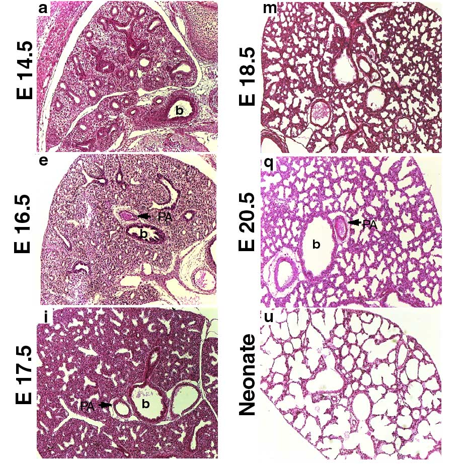 | 922 × 922 (239 KB) | S8600021 (talk | contribs) | ==Histological analyses of Mouse Lungs at Various Embryonic Stages== Lung sections stained with H and E, and taken at various gestational stages as indicated. Images are at lower magnification (10X) The images are representative sections from wild-type |
You cannot overwrite this file.
File usage
There are no pages that use this file.