File:Mouse hypothalamus SHH expression 01.jpg

Original file (600 × 902 pixels, file size: 115 KB, MIME type: image/jpeg)
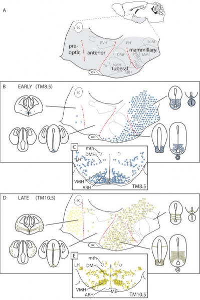
Mouse Hypothalamus Sonic hedgehog Expression
Distribution of sonic hedgehog Shh-lineage cells from the different progenitor domains in the mouse adult hypothalamus.
(A) Adult mouse brain in sagittal view showing the hypothalamus (shaded gray).
(B, D) Summary of distribution of cells fate-mapped at early and late stages. Magnified view of the shaded area showing the transverse hypothalamic subdivisions and some of the largest nuclei and Shh-lineage progenitor domains in the E12.5 neuroepithelium (small profiles; see also Figure 2). In the schematic of the adult hypothalamus, closed circles indicate neurons, open circles indicate astrocytes, and stars indicate tanycytes.
(C, E) Transverse section through the adult tuberal hypothalamus.
Legend
|
|
Reference
Alvarez-Bolado G, Paul FA & Blaess S. (2012). Sonic hedgehog lineage in the mouse hypothalamus: from progenitor domains to hypothalamic regions. Neural Dev , 7, 4. PMID: 22264356 DOI.
Copyright
© 2012 Alvarez-Bolado et al.; licensee BioMed Central Ltd
This is an open access article distributed under the terms of the Creative Commons Attribution License (http://creativecommons.org/licenses/by/2.0), which permits unrestricted use, distribution, and reproduction in any medium, provided the original work is properly cited.
Figure 8. 1749-8104-7-4-8.jpg
Cite this page: Hill, M.A. (2024, April 27) Embryology Mouse hypothalamus SHH expression 01.jpg. Retrieved from https://embryology.med.unsw.edu.au/embryology/index.php/File:Mouse_hypothalamus_SHH_expression_01.jpg
- © Dr Mark Hill 2024, UNSW Embryology ISBN: 978 0 7334 2609 4 - UNSW CRICOS Provider Code No. 00098G
File history
Click on a date/time to view the file as it appeared at that time.
| Date/Time | Thumbnail | Dimensions | User | Comment | |
|---|---|---|---|---|---|
| current | 13:38, 13 May 2012 |  | 600 × 902 (115 KB) | Z8600021 (talk | contribs) | ==Mouse Hypothalamus SHH Expression== Distribution of Shh-lineage cells from the different progenitor domains in the mouse adult hypothalamus. (A) Adult mouse brain in sagittal view showing the hypothalamus (shaded gray). (B, D) Summary of distribution o |
You cannot overwrite this file.
File usage
The following page uses this file: