File:Mouse fragilis2 expression.jpg
Original file (600 × 675 pixels, file size: 60 KB, MIME type: image/jpeg)
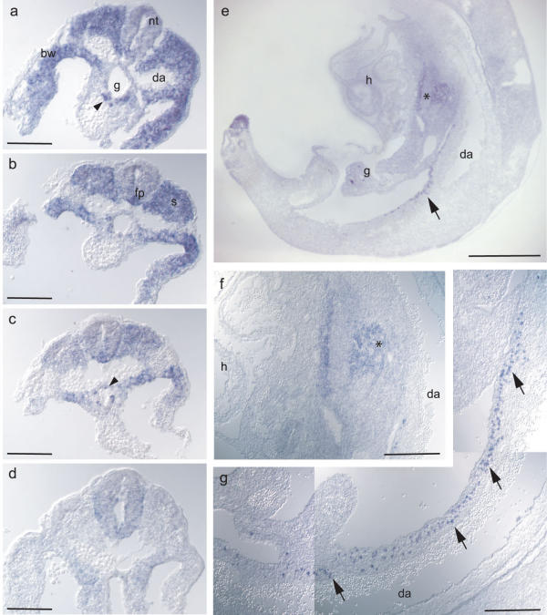
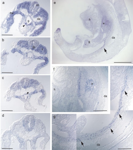
Expression analysis of fragilis2 by in situ hybridisation on sections.
(a-d) transverse sections through the caudal region of an embryo at E9.5 (approx. 25 somites) at progressively rostral levels. At most caudal levels, fragilis2 expression is seen in cells of the neural tube, in the presomitic mesoderm, in single cells within the hindgut (arrowhead) and in the body wall.
(b) staining at approx. 23rd somite level is present within the forming somite, the body wall mesoderm and cells within the hindgut as well as the floorplate.
(c) at approx. 21st somite level, expression in the differentiating somites is reduced, while cells in the floor plate and within the hindgut remain fragilis2 mRNA positive.
(d) at approx. the 13th somite level, fragilis2 expression is absent from the somatic mesoderm as well as the neural tube.
(e) sagittal section through an E10.5 embryo shows fragilis2 expression in developing lung tissue (asterix; higher magnification in f) and migrating cells along the hindgut anterior to the dorsal aorta (arrow).
(g) shows a magnified view of fragilis2 mRNA expressing, migrating cells.
da: dorsal aorta; fp: floor plate; g: gut; h: developing heart; nt: neural tube; s: somite; bw: body wall; scale bars: 150 μm (a-d); 1 mm (e); 400 μ m (f, g).
http://www.pubmedcentral.nih.gov/articlerender.fcgi?artid=153542&rendertype=figure&id=F4
The Fragilis interferon-inducible gene family of transmembrane proteins is associated with germ cell specification in mice UC Lange, M Saitou, PS Western, SC Barton, and MA Surani BMC Dev Biol. 2003; 3: 1. Published online 2003 March 19. doi: 10.1186/1471-213X-3-1. PMCID: PMC153542
BMC Dev Biol. 2003; 3: 1.
Published online 2003 March 19. doi: 10.1186/1471-213X-3-1.
Copyright © 2003 Lange et al; licensee BioMed Central Ltd. This is an Open Access article: verbatim copying and redistribution of this article are permitted in all media for any purpose, provided this notice is preserved along with the article's original URL.
File history
Click on a date/time to view the file as it appeared at that time.
| Date/Time | Thumbnail | Dimensions | User | Comment | |
|---|---|---|---|---|---|
| current | 17:24, 21 September 2009 | | 600 × 675 (60 KB) | S8600021 (talk | contribs) | Expression analysis of fragilis2 by in situ hybridisation on sections. (a-d) transverse sections through the caudal region of an embryo at E9.5 (approx. 25 somites) at progressively rostral levels. At most caudal levels, fragilis2 expression is seen in |
You cannot overwrite this file.
File usage
There are no pages that use this file.