File:Mouse follicle in vitro.jpg: Difference between revisions
No edit summary |
|||
| Line 1: | Line 1: | ||
== | ==In Vitro Grown Mouse Follicles== | ||
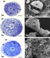
FCS follicles (panels a, b); FSH antral follicles (panels c, d); FSH non-antral follicles (panels e, f). The general appearance of cultured follicles is shown by LM (panels a, c, e) and SEM (panels b, d, f). | FCS follicles (panels a, b); FSH antral follicles (panels c, d); FSH non-antral follicles (panels e, f). The general appearance of cultured follicles is shown by LM (panels a, c, e) and SEM (panels b, d, f). | ||
| Line 7: | Line 7: | ||
Bar is: 30 μm (panel a); 100 μm (panels b, d); 50 μm (panels c, f); 45 μm (panel e). | Bar is: 30 μm (panel a); 100 μm (panels b, d); 50 μm (panels c, f); 45 μm (panel e). | ||
{{Mouse follicles in vitro}} | |||
==Reference== | ==Reference== | ||
| Line 15: | Line 19: | ||
This is an Open Access article distributed under the terms of the Creative Commons Attribution License (http://creativecommons.org/licenses/by/2.0), which permits unrestricted use, distribution, and reproduction in any medium, provided the original work is properly cited. | This is an Open Access article distributed under the terms of the Creative Commons Attribution License (http://creativecommons.org/licenses/by/2.0), which permits unrestricted use, distribution, and reproduction in any medium, provided the original work is properly cited. | ||
Original file name: Figure 1. 1477-7827-9-3-1.jpg | |||
[[Category:Mouse]] [[Category:Oocyte]] [[Category:Electron Micrograph]] | [[Category:Mouse]] [[Category:Oocyte]] [[Category:Electron Micrograph]] | ||
Latest revision as of 17:22, 12 February 2012
In Vitro Grown Mouse Follicles
FCS follicles (panels a, b); FSH antral follicles (panels c, d); FSH non-antral follicles (panels e, f). The general appearance of cultured follicles is shown by LM (panels a, c, e) and SEM (panels b, d, f).
Panels a-f, O: oocyte; GC: granulosa cells; TC: theca cells. Panels a, c, e, ZP: zona pellucida; b: basement membrane. Panel c, asterisks: fluid-filled spaces.
Bar is: 30 μm (panel a); 100 μm (panels b, d); 50 μm (panels c, f); 45 μm (panel e).
- Image Links: All images | follicle LM | follicle SEM | non-antral follicle LM | non-antral follicle SEM | antral follicle LM | antral follicle SEM | Oocyte Development | Mouse Development
Reference
<pubmed>21232101</pubmed>| PMC3033320 | Reprod Biol Endocrinol.
© 2011 Nottola et al; licensee BioMed Central Ltd.
This is an Open Access article distributed under the terms of the Creative Commons Attribution License (http://creativecommons.org/licenses/by/2.0), which permits unrestricted use, distribution, and reproduction in any medium, provided the original work is properly cited.
Original file name: Figure 1. 1477-7827-9-3-1.jpg
File history
Click on a date/time to view the file as it appeared at that time.
| Date/Time | Thumbnail | Dimensions | User | Comment | |
|---|---|---|---|---|---|
| current | 23:27, 23 February 2011 |  | 600 × 701 (170 KB) | S8600021 (talk | contribs) | ==General appearance of in vitro grown follicles== FCS follicles (panels a, b); FSH antral follicles (panels c, d); FSH non-antral follicles (panels e, f). The general appearance of cultured follicles is shown by LM (panels a, c, e) and SEM (panels b, d, |
You cannot overwrite this file.
File usage
The following page uses this file: