File:Mouse eye TGF-beta model.jpg
Original file (1,000 × 519 pixels, file size: 88 KB, MIME type: image/jpeg)
Mouse Eye TGF-beta Model
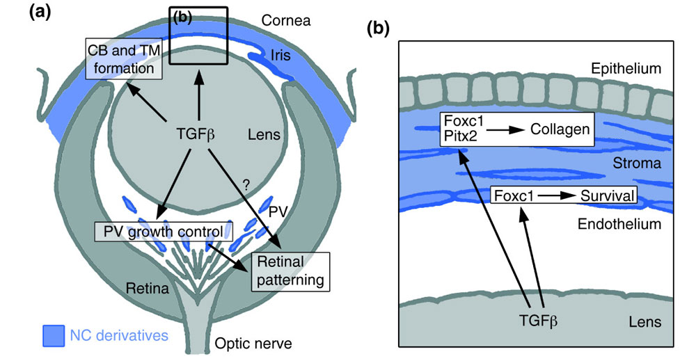
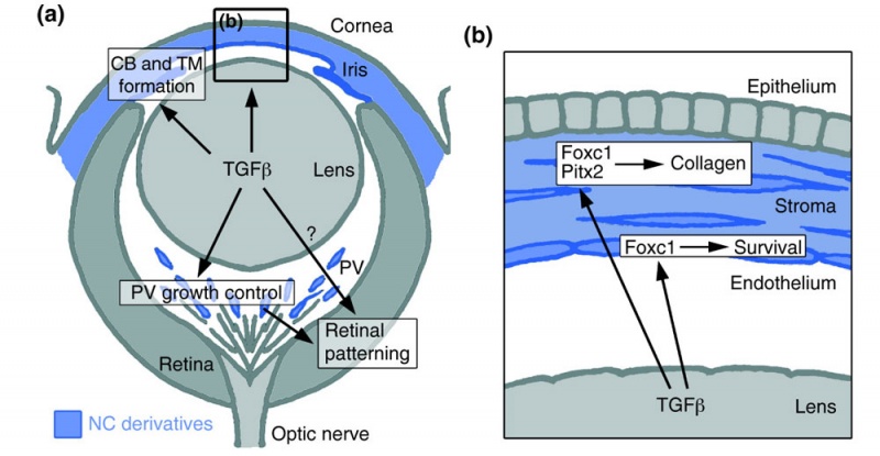
Summary of the TGFβ-dependent development of anterior and posterior ocular structures.
(a) NC-derived cells (blue) contribute to structures of the anterior eye segment and the primary vitreous (PV). TGFβ signaling is involved in the formation of the ciliary body (CB) and the trabecular meshwork (TM), and in control of PV growth. Moreover, normal PV development and/or TGFβ signaling are important for correct retinal patterning.
(b) In the cornea, prospective stromal keratocytes and endothelial cells are of NC origin. Here, TGFβ signaling is needed for the expression of the transcription factors Foxc1 and Pitx2 and for normal differentiation of NC-derived cells into collagen-synthesizing stromal keratocytes. Moreover, in forming corneal endothelial cells (and in the TM), expression of Foxc1 and cell survival requires TGFβ signalling.
Figure 9. http://jbiol.com/content/4/3/11/figure/F9
Reference
<pubmed>16403239</pubmed>| J Biol.
Ittner et al. Journal of Biology 2005 4:11 doi:10.1186/jbiol29
© 2005 Ittner et al.; licensee BioMed Central Ltd. This is an open access article distributed under the terms of the Creative Commons Attribution License (http://creativecommons.org/licenses/by/2.0), which permits unrestricted use, distribution, and reproduction in any medium, provided the original work is properly cited.
File history
Click on a date/time to view the file as it appeared at that time.
| Date/Time | Thumbnail | Dimensions | User | Comment | |
|---|---|---|---|---|---|
| current | 18:32, 30 August 2011 | | 1,000 × 519 (88 KB) | S8600021 (talk | contribs) | ==Mouse eye TGF-beta model== Summary of the TGFβ-dependent development of anterior and posterior ocular structures. (a) NC-derived cells (blue) contribute to structures of the anterior eye segment and the primary vitreous (PV). TGFβ signaling is invo |
You cannot overwrite this file.
File usage
The following 5 pages use this file: