File:Mouse eye E18.jpg: Difference between revisions
(==Mouse Eye (E18)== Toluidine blue staining of semi-thin sagittal sections of eyes at E18. {{Mouse eye neural crest links}} ===Reference=== <pubmed>16403239</pubmed>| [http://jbiol.com/content/4/3/11 J Biol.] ====Copyright==== © 2005 Ittner et...) |
|||
| (One intermediate revision by the same user not shown) | |||
| Line 2: | Line 2: | ||
Toluidine blue staining of semi-thin sagittal sections of eyes at E18. | Toluidine blue staining of semi-thin sagittal sections of eyes at E18. | ||
Compound ocular anomalies in Tgfbr2-mutant mice. (a) Toluidine blue staining of semi-thin sagittal sections of eyes at E18 reveals a smaller size with no anterior chamber and an infiltration of cells behind the lens in Tgfbr2-mutant embryos as compared with control embryos. Boxes indicate magnified regions shown in the other panels; scale bars represent 250 μm. | |||
* '''b''' - corneal stroma | |||
* '''c''' - Structures of the forming chamber angle, including the trabecular meshwork (black arrowhead) | |||
* '''d''' - primary vitreous consists of loosely arranged vessels of the hyaloid vascular system (arrows). | |||
* '''e''' - retina displays typical patterning, with clear separation into an inner layer (IRL) and an outer layer (ORL). | |||
| Line 18: | Line 24: | ||
Ittner et al. Journal of Biology 2005 4:11 doi:10.1186/jbiol29 | Ittner et al. Journal of Biology 2005 4:11 doi:10.1186/jbiol29 | ||
Original file name: Jbiol29-3.jpg | Original file name: Jbiol29-3.jpg Original figure a-e cropped, resized and relabeled. | ||
[[Category:Mouse]] [[Category:Neural Crest]] [[Category:Vision]] [[Category:Cornea]] | [[Category:Mouse]] [[Category:Neural Crest]] [[Category:Vision]] [[Category:Cornea]] | ||
[[Category:Mouse E18]] | [[Category:Mouse E18]] | ||
Latest revision as of 15:44, 30 August 2014
Mouse Eye (E18)
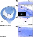
Toluidine blue staining of semi-thin sagittal sections of eyes at E18.
Compound ocular anomalies in Tgfbr2-mutant mice. (a) Toluidine blue staining of semi-thin sagittal sections of eyes at E18 reveals a smaller size with no anterior chamber and an infiltration of cells behind the lens in Tgfbr2-mutant embryos as compared with control embryos. Boxes indicate magnified regions shown in the other panels; scale bars represent 250 μm.
- b - corneal stroma
- c - Structures of the forming chamber angle, including the trabecular meshwork (black arrowhead)
- d - primary vitreous consists of loosely arranged vessels of the hyaloid vascular system (arrows).
- e - retina displays typical patterning, with clear separation into an inner layer (IRL) and an outer layer (ORL).
- Links: Image - Mouse eye neural crest | Image - cornea and anterior chamber | Image - cornea | Image - Mouse eye TGF-beta model | Vision - Cornea Development | Vision Development | Neural Crest | Head Development
Reference
<pubmed>16403239</pubmed>| J Biol.
Copyright
© 2005 Ittner et al.; licensee BioMed Central Ltd. This is an open access article distributed under the terms of the Creative Commons Attribution License (http://creativecommons.org/licenses/by/2.0), which permits unrestricted use, distribution, and reproduction in any medium, provided the original work is properly cited.
Ittner et al. Journal of Biology 2005 4:11 doi:10.1186/jbiol29
Original file name: Jbiol29-3.jpg Original figure a-e cropped, resized and relabeled.
File history
Click on a date/time to view the file as it appeared at that time.
| Date/Time | Thumbnail | Dimensions | User | Comment | |
|---|---|---|---|---|---|
| current | 15:12, 30 August 2014 |  | 884 × 977 (169 KB) | Z8600021 (talk | contribs) | ==Mouse Eye (E18)== Toluidine blue staining of semi-thin sagittal sections of eyes at E18. {{Mouse eye neural crest links}} ===Reference=== <pubmed>16403239</pubmed>| [http://jbiol.com/content/4/3/11 J Biol.] ====Copyright==== © 2005 Ittner et... |
You cannot overwrite this file.
File usage
There are no pages that use this file.