File:Mouse CT axes E11.5.jpg: Difference between revisions
No edit summary |
No edit summary |
||
| Line 1: | Line 1: | ||
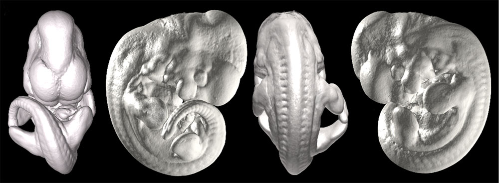
==Mouse Embryo Computed Tomography== | |||
Isosurface of E11.5 embryos scanned at 8-μm resolution. | |||
Comparison of Virtual and Paraffin Histology of an E11.5 Embryo Scanned at 8 μm | Comparison of Virtual and Paraffin Histology of an E11.5 Embryo Scanned at 8 μm | ||
(A) Isosurface renderings of the CT-scanned embryo. | :'''(A)''' Isosurface renderings of the CT-scanned embryo. | ||
:'''(B)''' Maximum intensity projection of the same embryo, with overlying places of section. | |||
( | :'''(C–E)''' Sagittal, coronal, and axial sections of an E11.5 littermate. | ||
( | :'''(F–H)''' Sagittal, coronal, and axial computed tomography sections of the embryo in panels (A) and (B), corresponding to the planes of section in panels (C–E). At low-power magnification, virtual and true paraffin histology demonstrate a similar level of detail. Scale bar indicates 400 μm. | ||
a, cardiac atrium; cv, cardinal vein; drg, dorsal root ganglia; fv, forebrain vesicle; liv, liver; nt, neural tube; v, cardiac ventricle; v4, fourth ventricle. | a, cardiac atrium; cv, cardinal vein; drg, dorsal root ganglia; fv, forebrain vesicle; liv, liver; nt, neural tube; v, cardiac ventricle; v4, fourth ventricle. | ||
| Line 14: | Line 19: | ||
Original File Name:journal.pgen.0020061.g001.tif | Original File Name:journal.pgen.0020061.g001.tif | ||
<pubmed>16683035</pubmed>| [http://www.plosgenetics.org/article/info:doi/10.1371/journal.pgen.0020061 PLoS Genetics] | |||
http://www.plosgenetics.org/article/info:doi/10.1371/journal.pgen.0020061 | |||
Citation: Johnson JT, Hansen MS, Wu I, Healy LJ, Johnson CR, et al. (2006) Virtual Histology of Transgenic Mouse Embryos for High-Throughput Phenotyping. PLoS Genet 2(4): e61. doi:10.1371/journal.pgen.0020061 | Citation: Johnson JT, Hansen MS, Wu I, Healy LJ, Johnson CR, et al. (2006) Virtual Histology of Transgenic Mouse Embryos for High-Throughput Phenotyping. PLoS Genet 2(4): e61. doi:10.1371/journal.pgen.0020061 | ||
Revision as of 10:00, 17 August 2010
Mouse Embryo Computed Tomography
Isosurface of E11.5 embryos scanned at 8-μm resolution.
Comparison of Virtual and Paraffin Histology of an E11.5 Embryo Scanned at 8 μm
- (A) Isosurface renderings of the CT-scanned embryo.
- (B) Maximum intensity projection of the same embryo, with overlying places of section.
- (C–E) Sagittal, coronal, and axial sections of an E11.5 littermate.
- (F–H) Sagittal, coronal, and axial computed tomography sections of the embryo in panels (A) and (B), corresponding to the planes of section in panels (C–E). At low-power magnification, virtual and true paraffin histology demonstrate a similar level of detail. Scale bar indicates 400 μm.
a, cardiac atrium; cv, cardinal vein; drg, dorsal root ganglia; fv, forebrain vesicle; liv, liver; nt, neural tube; v, cardiac ventricle; v4, fourth ventricle.
doi:10.1371/journal.pgen.0020061.g001
Original File Name:journal.pgen.0020061.g001.tif
<pubmed>16683035</pubmed>| PLoS Genetics
Citation: Johnson JT, Hansen MS, Wu I, Healy LJ, Johnson CR, et al. (2006) Virtual Histology of Transgenic Mouse Embryos for High-Throughput Phenotyping. PLoS Genet 2(4): e61. doi:10.1371/journal.pgen.0020061
Editor: Wayne Frankel, The Jackson Laboratory, United States of America
Received: January 23, 2006; Accepted: March 13, 2006; Published: April 28, 2006
Copyright: © 2006 Johnson et al. This is an open-access article distributed under the terms of the Creative Commons Attribution License, which permits unrestricted use, distribution, and reproduction in any medium, provided the original author and source are credited.
File history
Click on a date/time to view the file as it appeared at that time.
| Date/Time | Thumbnail | Dimensions | User | Comment | |
|---|---|---|---|---|---|
| current | 01:03, 16 April 2010 | 1,000 × 367 (68 KB) | S8600021 (talk | contribs) | Comparison of Virtual and Paraffin Histology of an E11.5 Embryo Scanned at 8 μm (A) Isosurface renderings of the CT-scanned embryo. (B) Maximum intensity projection of the same embryo, with overlying places of section. (C–E) Sagittal, coronal, and a |
You cannot overwrite this file.
File usage
The following 5 pages use this file: