File:Mouse-spermatozoa SLY protein.jpg

Original file (637 × 767 pixels, file size: 352 KB, MIME type: image/jpeg)
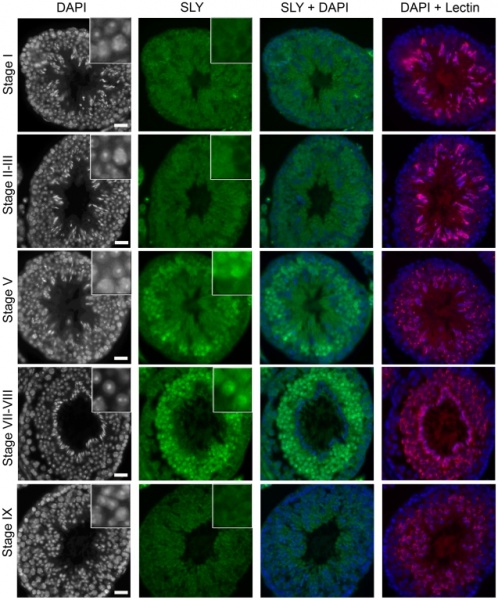
SLY protein is located in the spermatid nuclei in a stage-specific manner.
Detection of SLY protein (green) by immunofluorescence in wild-type testis sections. DAPI (white, or blue in the merged picture) was used to stain nuclei, and lectin-PNA (red) was used to stain acrosomes in order to determine tubule stage.
The inset in the upper-right corner represents a 3.4× magnification. Pictures were taken using the same image capture parameters.
Scale bars indicate 20 µm. See Figure S9 for a complete panel.
Figure 7 Pbio.1000244.g007.jpg
http://www.ncbi.nlm.nih.gov/pmc/articles/PMC2770110/?tool=pmcentrez
- "SLY protein colocalizes with the X and Y chromatin in spermatids of normal males, and Sly deficiency leads to defective repressive marks on the sex chromatin, such as reduced levels of the heterochromatin protein CBX1 and of histone H3 methylated at lysine 9."
PLoS Biol. 2009 November; 7(11): e1000244.
Published online 2009 November 17. doi: 10.1371/journal.pbio.1000244.
Copyright Cocquet et al. This is an open-access article distributed under the terms of the Creative Commons Attribution License, which permits unrestricted use, distribution, and reproduction in any medium, provided the original author and source are credited.
File history
Click on a date/time to view the file as it appeared at that time.
| Date/Time | Thumbnail | Dimensions | User | Comment | |
|---|---|---|---|---|---|
| current | 14:40, 5 April 2010 | | 637 × 767 (352 KB) | S8600021 (talk | contribs) | SLY protein is located in the spermatid nuclei in a stage-specific manner. Detection of SLY protein (green) by immunofluorescence in wild-type testis sections. DAPI (white, or blue in the merged picture) was used to stain nuclei, and lectin-PNA (red) was |
You cannot overwrite this file.
File usage
There are no pages that use this file.