File:Mouse- spermatozoa EM and diagram.jpg: Difference between revisions
(==Mouse Spermatozoa EM and Diagram== The Spermatozoon: the spermatozoon is made up of two main regions, the head and the tail. The anterior portion of the head is covered by the acrosomal cap and the head is joined to the tail by the connecting piece. The) |
|||
| Line 7: | Line 7: | ||
Original File Name: Figure 1 http://www.ncbi.nlm.nih.gov/pmc/articles/PMC2816191/figure/DMP032F1/ (extracted from full figure) | Original File Name: Figure 1 http://www.ncbi.nlm.nih.gov/pmc/articles/PMC2816191/figure/DMP032F1/ (extracted from full figure) | ||
==Reference== | ===Reference=== | ||
<pubmed>19758979</pubmed>| [http://www.ncbi.nlm.nih.gov/pmc/articles/PMC2816191 PMC2816191] | [http://humupd.oxfordjournals.org/content/16/2/205.long Hum Reprod Update.] | <pubmed>19758979</pubmed>| [http://www.ncbi.nlm.nih.gov/pmc/articles/PMC2816191 PMC2816191] | [http://humupd.oxfordjournals.org/content/16/2/205.long Hum Reprod Update.] | ||
| Line 15: | Line 15: | ||
Hum Reprod Update. 2010 Mar–Apr; 16(2): 205–224. | Hum Reprod Update. 2010 Mar–Apr; 16(2): 205–224. | ||
Published online 2009 September 15. doi: 10.1093/humupd/dmp032. | Published online 2009 September 15. doi: 10.1093/humupd/dmp032. | ||
This is an Open Access article distributed under the terms of the Creative Commons Attribution Non-Commercial License (http://creativecommons.org/licenses/by-nc/2.5/uk/) which permits unrestricted non-commercial use, distribution, and reproduction in any medium, provided the original work is properly cited. | This is an Open Access article distributed under the terms of the Creative Commons Attribution Non-Commercial License (http://creativecommons.org/licenses/by-nc/2.5/uk/) which permits unrestricted non-commercial use, distribution, and reproduction in any medium, provided the original work is properly cited. | ||
[[Category:Mouse]] [[Category:Spermatozoa]] [[Category:Cartoon]] | [[Category:Mouse]] [[Category:Spermatozoa]] [[Category:Cartoon]] | ||
Revision as of 10:01, 18 February 2012
Mouse Spermatozoa EM and Diagram
The Spermatozoon: the spermatozoon is made up of two main regions, the head and the tail. The anterior portion of the head is covered by the acrosomal cap and the head is joined to the tail by the connecting piece. The tail is divided into three regions: the midpiece; principal piece; and the end-piece.
The electron micrographs showing cross-sections (not to scale) of each region highlights the main components of the tail structure: the axoneme; outer dense fibers (ODF); and the mitochondrial sheath (midpiece) and fibrous sheath (FS) (principal piece). The end-piece consists solely of the axoneme and plasma membrane.
Original File Name: Figure 1 http://www.ncbi.nlm.nih.gov/pmc/articles/PMC2816191/figure/DMP032F1/ (extracted from full figure)
Reference
<pubmed>19758979</pubmed>| PMC2816191 | Hum Reprod Update.
From: Hum Reprod Update. 2010 Mar–Apr; 16(2): 205–224. Published online 2009 September 15. doi: 10.1093/humupd/dmp032.
This is an Open Access article distributed under the terms of the Creative Commons Attribution Non-Commercial License (http://creativecommons.org/licenses/by-nc/2.5/uk/) which permits unrestricted non-commercial use, distribution, and reproduction in any medium, provided the original work is properly cited.
File history
Click on a date/time to view the file as it appeared at that time.
| Date/Time | Thumbnail | Dimensions | User | Comment | |
|---|---|---|---|---|---|
| current | 12:52, 24 October 2010 | 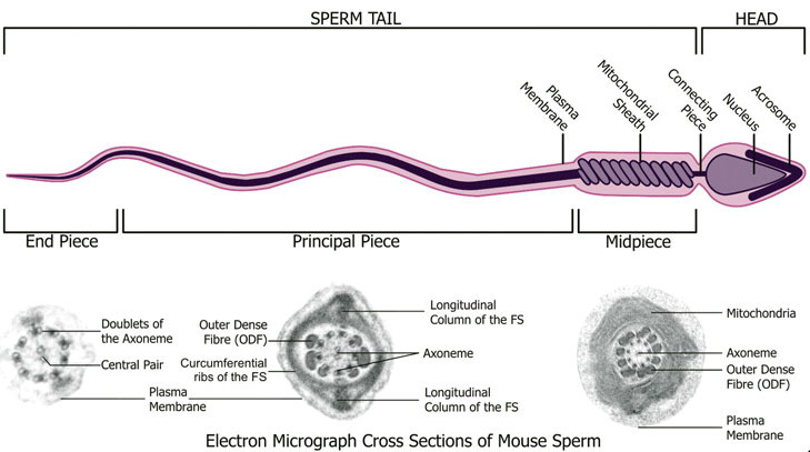 | 729 × 407 (49 KB) | S8600021 (talk | contribs) | ==Mouse Spermatozoa EM and Diagram== The Spermatozoon: the spermatozoon is made up of two main regions, the head and the tail. The anterior portion of the head is covered by the acrosomal cap and the head is joined to the tail by the connecting piece. The |
You cannot overwrite this file.
File usage
The following 5 pages use this file: