File:Morton1949 plate02.jpg: Difference between revisions
From Embryology
mNo edit summary |
mNo edit summary |
||
| Line 1: | Line 1: | ||
==Plate 2== | ==Plate 2== | ||
Fig.4. The embryonic rudiment. The distal expansion of the yolk-sac is not figured. Sect.81. x140. | [[:File:Morton1949 fig04.jpg|'''Fig. 4''']]. The embryonic rudiment. The distal expansion of the yolk-sac is not figured. Sect.81. x140. | ||
Fig. 5. The opening of the duct part into the distal expansion of the yolk-sac which shows its thin ab-embryonic wall below. Sect.75. x95. | [[:File:Morton1949 fig05.jpg|'''Fig. 5''']]. The opening of the duct part into the distal expansion of the yolk-sac which shows its thin ab-embryonic wall below. Sect.75. x95. | ||
Fig. 6. The Macafee ovum in situ. Diagnostic section. x 65. | [[:File:Morton1949 fig06.jpg|'''Fig. 6''']]. The Macafee ovum in situ. Diagnostic section. x 65. | ||
<gallery> | |||
File:Morton1949 fig04.jpg|Fig. 4 | |||
File:Morton1949 fig05.jpg|Fig. 5 | |||
File:Morton1949 fig06.jpg|Fig. 6 | |||
</gallery> | |||
{{Historic Disclaimer}} | {{Historic Disclaimer}} | ||
Latest revision as of 10:56, 11 August 2017
Plate 2
Fig. 4. The embryonic rudiment. The distal expansion of the yolk-sac is not figured. Sect.81. x140.
Fig. 5. The opening of the duct part into the distal expansion of the yolk-sac which shows its thin ab-embryonic wall below. Sect.75. x95.
Fig. 6. The Macafee ovum in situ. Diagnostic section. x 65.
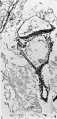
Fig. 4
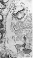
Fig. 5
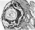
Fig. 6


| Historic Disclaimer - information about historic embryology pages |
|---|
 |
Cite this page: Hill, M.A. (2024, May 19) Embryology Morton1949 plate02.jpg. Retrieved from https://embryology.med.unsw.edu.au/embryology/index.php/File:Morton1949_plate02.jpg
- © Dr Mark Hill 2024, UNSW Embryology ISBN: 978 0 7334 2609 4 - UNSW CRICOS Provider Code No. 00098G
File history
Click on a date/time to view the file as it appeared at that time.
| Date/Time | Thumbnail | Dimensions | User | Comment | |
|---|---|---|---|---|---|
| current | 20:09, 9 August 2015 |  | 1,598 × 2,499 (1 MB) | Z8600021 (talk | contribs) | ==Plate 1== Fig. 1. The ovum in 8itu. Sect. 81. x 38. Fig. 2. The duct part of the yolk-sac. The distal end is inferior. Sect. 77. x 375. Fig. 3. The distal expansion of the yolk-sac. The end of the duct just appears above, on the left. Sect.77. x375. |
You cannot overwrite this file.
File usage
The following page uses this file: