File:Morton1949 plate01.jpg
From Embryology

Size of this preview: 435 × 599 pixels. Other resolution: 1,784 × 2,457 pixels.
Original file (1,784 × 2,457 pixels, file size: 975 KB, MIME type: image/jpeg)
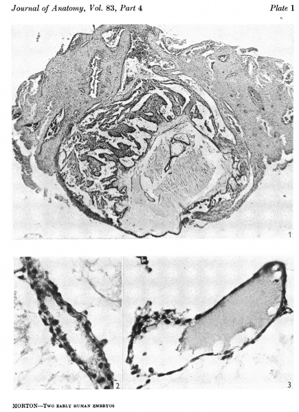
Plate 1
Fig. 1. The ovum in 8itu. Sect. 81. x 38.
Fig. 2. The duct part of the yolk-sac. The distal end is inferior. Sect. 77. x 375.
Fig. 3. The distal expansion of the yolk-sac. The end of the duct just appears above, on the left. Sect.77. x375.
File history
Click on a date/time to view the file as it appeared at that time.
| Date/Time | Thumbnail | Dimensions | User | Comment | |
|---|---|---|---|---|---|
| current | 20:10, 9 August 2015 |  | 1,784 × 2,457 (975 KB) | Z8600021 (talk | contribs) |
You cannot overwrite this file.
File usage
The following page uses this file: