File:Moffatt1957 plate02.jpg
Original file (1,500 × 1,891 pixels, file size: 865 KB, MIME type: image/jpeg)
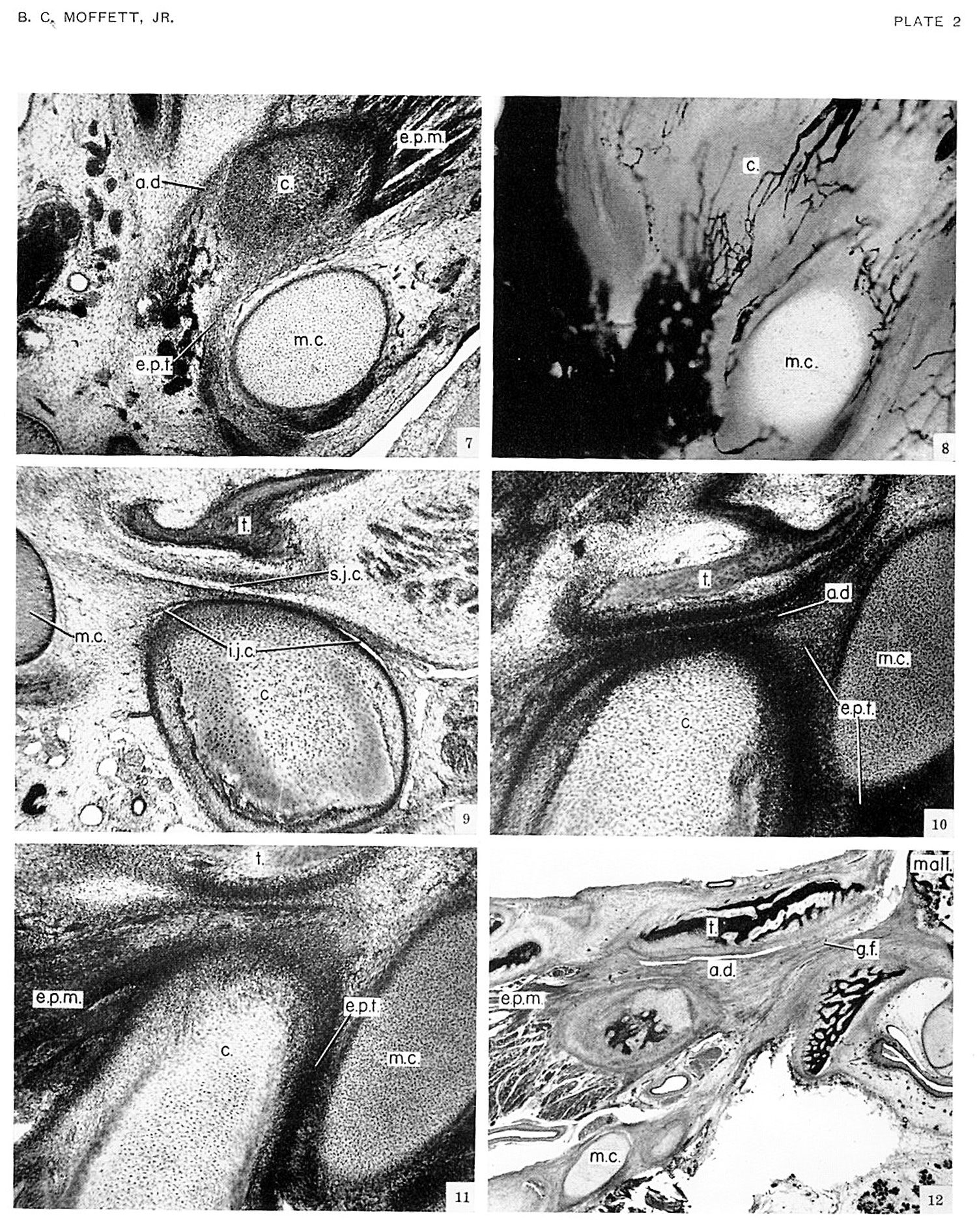
Plate 2
Fig 7 - fetal (no. 3990 54 mm) The attachment of the developing disk to Meckel’s cartilage can be seen also in superior view by looking down on a transverse or horizontal section through the joint. Here the tendon of the external pterygoid muscle passes over the medial side of the condyle and then turns, proceeding to the lateral side of Meckel’s cartilage, on its way to the head of the malleus. The contribution from the disk over the lateral part of the condyle is more readily visible a few sections higher, where it joins this tendon as a somewhat more deeply stained band of cells.
Fig 8 - fetal (no. 858 57.25 mm) At the level shown, these blood vessels give off branches which run anteriorly in the articular disk and on the ternal pterygoid tendon to anastomose with capillaries coming from the anterior surface of the joint. This capilry anastomosis, extending anteroposteriorly completely zross the articular disk, is shown in a horizontal section, in which re vascular system was injected with India ink. The level of section corresponds to that shown in figure 7, Id passes through the disk and pterygoid tendon immediately above the condyle.
Fig 9 - fetal (no. 6581 60 mm) Sagittal section the inferior joint cavity is present 1n the superior cavity is being formed. Development of joint capsule. The articular capsule of re temporomandibular joint is first recognizable in )-mm. fetuses in the form of faint cellular condensations along the medial and lateral sides of the joint, conecting the two skeletal components. The articular disk terges peripherally with these capsular condensations 1d thus obtains an attachment to the temporal bone 1d mandible. The joint is still not bounded anteroasteriorly by a capsule in the 75-mm. fetus. Before the irmation of the capsule posterior to the joint, the articular disk transfers its attachment from Meckel’s cartilage in the temporal bone and mandible. This transfer results from a narrowing of the Glaserian fissure and an approximation of Meckel’s cartilage to the posterior part of the neck of the condyle.
Fig 10 - fetal (no. 1455 78.5 mm) An early phase in the trans fer to the temporal bone is seen in a sagittal section through the lateral portion of the temporamandibular joint. The articular disk here extends posteriorly, following the contours of the zygomatic process of the temporal bone, a relation which results from the approximation of the two articular elements and from the persisting attachment of the disk to Meckel’s cartilage. As the articular disk accompanies Meckel’s cartilage through the Glaserian fissure into the middle ear, the disk comes in contact with the anterior wall of the fissure. At this point the cells of the disk are becoming attached to the temporal bone. This attachment limits the superior joint cavity posteriorly and forms the superior attachment of the developing articular capsule on the anterior lip of the Glaserian fissure posterior to the temporomandibular jolnt.
Fig 11 - The inferior attachment of the disk and articular capsule on the posterior surface of the neck of the condyle results from a similar transfer involving the external pterygoid tendon, the mandible, and Meckel’s cartilage. The external pterygoid tendon is visible on the posterior surface of the condyle and again just inferior to Meckel’s cartilage as the tendon continues toward its insertion on the malleus. Further medially in a sagittal section through the middle of the condyle in the same specimen the tendon forms an attachment to the mandible. Here the external pterygoid tendon passes through a narrow space between the mandible and Meckel’s cartilage and appears to be attaching itself to the neck of the condyle. From this point, cells are seen extending superiorly toward the attachment of the disk on the temporal bone. At this stage, however, there is not a complete capsule posterior to the joint; the development of the capsule in this region proceeds very slowly.
Fig 12 - fetal (no. 625 220 mm) The attachments of the disk to the temporal bone and condyle are shown in sagittal section through the joint. This section passes through the medial part of the condyle. The external pterygoid muscle is seen merging with the anterior part of the disk. The Glaserian fissure is still open.
| Historic Disclaimer - information about historic embryology pages |
|---|
 |
Reference
Moffatt BC. The prenatal development of the human temporomandibular joint. (1957) Carnegie Instn. Wash. Publ. 611, Contrib. Embryol., 36: .
Cite this page: Hill, M.A. (2024, April 27) Embryology Moffatt1957 plate02.jpg. Retrieved from https://embryology.med.unsw.edu.au/embryology/index.php/File:Moffatt1957_plate02.jpg
- © Dr Mark Hill 2024, UNSW Embryology ISBN: 978 0 7334 2609 4 - UNSW CRICOS Provider Code No. 00098G
File history
Click on a date/time to view the file as it appeared at that time.
| Date/Time | Thumbnail | Dimensions | User | Comment | |
|---|---|---|---|---|---|
| current | 18:55, 30 October 2016 | | 1,500 × 1,891 (865 KB) | Z8600021 (talk | contribs) |
You cannot overwrite this file.
File usage
The following page uses this file: