File:Model digit origin.png
Model_digit_origin.png (599 × 505 pixels, file size: 128 KB, MIME type: image/png)
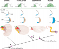
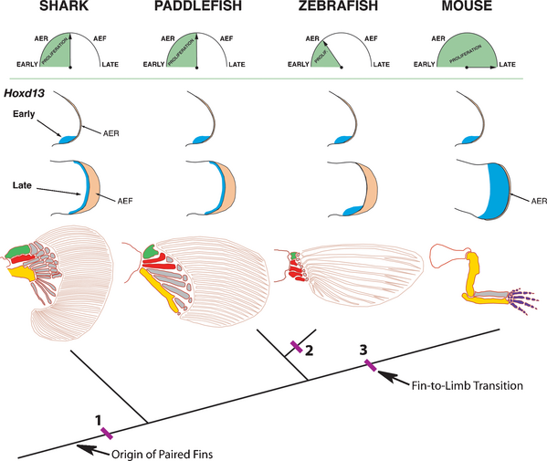
Model for the origin of digits by temporal extension of distal Hoxd13 expression
Tree shows phylogenetic relationships of shark, paddlefish, zebrafish and mouse. Top row shows hypothetical timing for the transition of the apical ectodermal ridge (AER) to apical ectodermal fold (AEF); note that the mouse maintains an AER and does not form an AEF.
Green shading represents proliferative period for endoskeletal progenitor cells.
Middle rows show Hoxd13 expression domains (blue) at early and late stages of fin and limb bud outgrowth. AER and AEF are shaded orange.
Bottom row shows pectoral appendicular skeleton for each taxon. Endoskeletal bones are shaded as follows: green, propterygium; red, mesopterygium, yellow, metapterygium. Dermal fin rays are shown as unshaded elements within fin blade.
The model suggests that a second phase of distal Hoxd13 expression was present in the paired fins of the common ancestor of chondrichthyans and osteichthyans (at position 1), and that loss of the distal Hoxd13 domain in teleosts (position 2) and its spatial expansion in tetrapods (position 3) may have been associated with temporal modulation of endoskeletal progenitor cell proliferation. Early conversion of the AER to an AEF would be expected to truncate or eliminate phase II expression of Hoxd13 and reduce the fin endoskeleton, as seen in zebrafish, whereas prolonged signaling by the AER would be expected to extend Phase II and expand the Hoxd13 domain, giving rise to digits in the tetrapod lineage.
Clock model after [25]; skeletal patterns after [15], [31], [40], [74].
Reference
Freitas R, Zhang G & Cohn MJ. (2007). Biphasic Hoxd gene expression in shark paired fins reveals an ancient origin of the distal limb domain. PLoS ONE , 2, e754. PMID: 17710153 DOI.
Citation: Freitas R, Zhang G, Cohn MJ (2007) Biphasic Hoxd Gene Expression in Shark Paired Fins Reveals an Ancient Origin of the Distal Limb Domain. PLoS ONE 2(8): e754. doi:10.1371/journal.pone.0000754
Copyright
© 2007 Freitas et al. This is an open-access article distributed under the terms of the Creative Commons Attribution License, which permits unrestricted use, distribution, and reproduction in any medium, provided the original author and source are credited.
Journal.pone.0000754.g005.png
http://www.plosone.org/article/info%3Adoi%2F10.1371%2Fjournal.pone.0000754
Cite this page: Hill, M.A. (2024, April 28) Embryology Model digit origin.png. Retrieved from https://embryology.med.unsw.edu.au/embryology/index.php/File:Model_digit_origin.png
- © Dr Mark Hill 2024, UNSW Embryology ISBN: 978 0 7334 2609 4 - UNSW CRICOS Provider Code No. 00098G
File history
Click on a date/time to view the file as it appeared at that time.
| Date/Time | Thumbnail | Dimensions | User | Comment | |
|---|---|---|---|---|---|
| current | 19:59, 14 September 2009 |  | 599 × 505 (128 KB) | S8600021 (talk | contribs) | Model for the origin of digits by temporal extension of distal Hoxd13 expression. Tree shows phylogenetic relationships of shark, paddlefish, zebrafish and mouse. Top row shows hypothetical timing for the transition of the apical ectodermal ridge (AER) t |
You cannot overwrite this file.
File usage
The following page uses this file:
