File:Minot1904 fig01.jpg
Minot1904_fig01.jpg (800 × 555 pixels, file size: 100 KB, MIME type: image/jpeg)
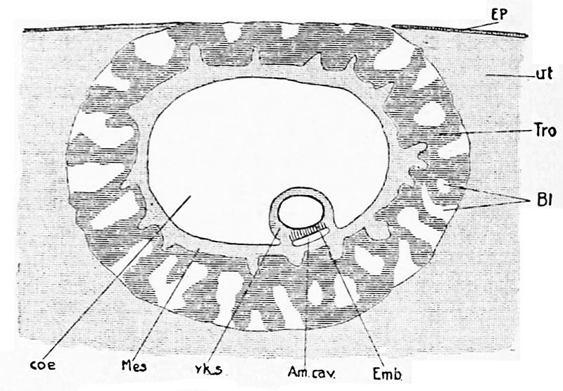
Explanation of diagram Based on Peters' Ovum
Ep, uterine epithelium; Ut, mucous membrane (decidua) of uterus; Tro, trophoderm ; Bl, spaces formed by the degeneration of the trophoderm ; maternal blood enters these spaces from the decidual bloodvessels ; Emb, embryonic shield; Am. cav, amniotic cavity; Yk.s, yolk sack, the entodermal lining of which is indicated by a heavy black line ; Mes, chorionic mesoderm ; Coe, extraembryonic celom.
Online Editor - O'Rahilly R. and Müller F. Developmental Stages in Human Embryos. Contrib. Embryol., Carnegie Inst. Wash. 637 (1987).
- "Peters Ovum described in a monograph by Peters (1899). Autopsy. A famous embryo, for long the youngest known and the first to be described in detail. Photomicrographs have since been published (Rossenbeck, 1923, plate 42; Odgers, 1937, plate 2, fig. 2). The chorionic villi, some of which display a mesenchymal core, send cellular columns externally and these latter are beginning to form a cytotrophoblastic shell. Slight branching of villi (Krafka, 1941). Chorionic cavity contains magma réticulé of Velpeau (Mall, 1916). Blood islands on umbilical vesicle. Chorion, 1.5 x 2 mm. Chorionic cavity, 1.6 x 0.9 x 0.8 mm; capacity, 0.7 mm3 (Odgers, 1937). Embryonic disc, 0.18 x 0.24 mm (Krafka, 1941). The basement membrane (Hensen’s membrane prima) of the epiblast was noted by Graf Spee. Allantoic diverticulum and primitive streak uncertain. Presumed age, 13 days (Krafka,1941). A tabulation of normal human embryos compiled from the literature prior to 1900 and from Mall’s own collection was published by Mall (1900, pp. 38-46). The least advanced specimen was the Peters embryo, and included in the list were 92 embryos of 0.19-32 mm, as well as 17 fetuses of 33-210 mm."
Reference
Minot CS. Implantation of the human ovum in the uterus. (1904) Trans. Am. Gynec. Soc., Philadelphia, 29: 395-402.
Cite this page: Hill, M.A. (2024, April 27) Embryology Minot1904 fig01.jpg. Retrieved from https://embryology.med.unsw.edu.au/embryology/index.php/File:Minot1904_fig01.jpg
- © Dr Mark Hill 2024, UNSW Embryology ISBN: 978 0 7334 2609 4 - UNSW CRICOS Provider Code No. 00098G
File history
Click on a date/time to view the file as it appeared at that time.
| Date/Time | Thumbnail | Dimensions | User | Comment | |
|---|---|---|---|---|---|
| current | 12:07, 9 February 2017 |  | 800 × 555 (100 KB) | Z8600021 (talk | contribs) | |
| 12:07, 9 February 2017 |  | 1,125 × 1,034 (249 KB) | Z8600021 (talk | contribs) | Transaction29ameruoft_0472.jpg |
You cannot overwrite this file.
File usage
The following page uses this file:
