File:Minot1883 fig01.jpg
From Embryology
No higher resolution available.
Minot1883_fig01.jpg (752 × 314 pixels, file size: 78 KB, MIME type: image/jpeg)
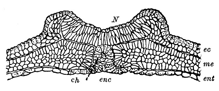
Frontal section through a frog ovum
Frontal section through a frog ovum in which the medullary ridges have begun to appear.
- Ent, entoderm
- enc, chordaentoblast
- ch, notochord
- me, mesoderm
- ec, ectoderm
- N, nervous system
Reference
Minot CS. Origin of mesoderm. (1883) Science 2(47);815-8 . PMID 17776824
Cite this page: Hill, M.A. (2024, April 27) Embryology Minot1883 fig01.jpg. Retrieved from https://embryology.med.unsw.edu.au/embryology/index.php/File:Minot1883_fig01.jpg
- © Dr Mark Hill 2024, UNSW Embryology ISBN: 978 0 7334 2609 4 - UNSW CRICOS Provider Code No. 00098G
File history
Click on a date/time to view the file as it appeared at that time.
| Date/Time | Thumbnail | Dimensions | User | Comment | |
|---|---|---|---|---|---|
| current | 17:56, 8 May 2016 | | 752 × 314 (78 KB) | Z8600021 (talk | contribs) | |
| 17:53, 8 May 2016 | 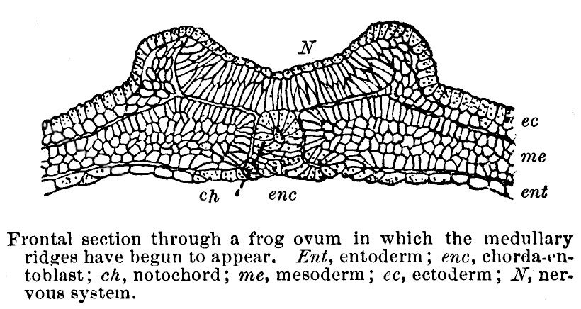 | 820 × 436 (111 KB) | Z8600021 (talk | contribs) |
You cannot overwrite this file.
File usage
The following page uses this file: