File:Microlissencephaly MRI-01.jpg
Original file (1,280 × 412 pixels, file size: 63 KB, MIME type: image/jpeg)
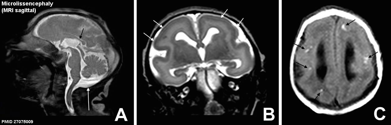
Microlissencephaly (MRI)
Sagittal T2 weighted image (A) and axial T2 weighted image (B) show moderate microcephalic brain, almost completely smooth cerebral surface with a thick cortex (short white arrows), and hypoplasia of corpus callosum (black arrow). In addition, the cisterna magna (long white arrow) is enlarged. Axial T1 weighted image (C) shows multiple hyperintense subcortical punctate dystrophic calcifications (black arrows)
- Links: Zika Virus | Magnetic Resonance
Reference
<pubmed>27075009</pubmed>| BMJ.
Copyright
This is an Open Access article distributed in accordance with the Creative Commons Attribution Non Commercial (CC BY-NC 3.0) license, which permits others to distribute, remix, adapt, build upon this work non-commercially, and license their derivative works on different terms, provided the original work is properly cited and the use is non-commercial. See: http://creativecommons.org/licenses/by-nc/3.0/.
Fig 4 relabelled.
Cite this page: Hill, M.A. (2024, May 7) Embryology Microlissencephaly MRI-01.jpg. Retrieved from https://embryology.med.unsw.edu.au/embryology/index.php/File:Microlissencephaly_MRI-01.jpg
- © Dr Mark Hill 2024, UNSW Embryology ISBN: 978 0 7334 2609 4 - UNSW CRICOS Provider Code No. 00098G
File history
Click on a date/time to view the file as it appeared at that time.
| Date/Time | Thumbnail | Dimensions | User | Comment | |
|---|---|---|---|---|---|
| current | 14:42, 28 April 2016 | 1,280 × 412 (63 KB) | Z8600021 (talk | contribs) | ==Microlissencephaly (MRI)== |
You cannot overwrite this file.
File usage
The following 2 pages use this file: