File:Meyer1920 fig27.jpg

Original file (352 × 651 pixels, file size: 30 KB, MIME type: image/jpeg)
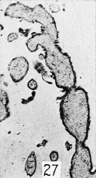
Figure 27
Not infrequently proliferation of the epithelium without increase in thickness may manifest itself in another way. The caliber of the villi in the earlicr stages of hydatiform degeneration sometimes does not increase much, no thickening of the proliferating epithelium is noticeable, and yet the latter shows marked proliferation. Under these circumstances, the borders of the villi and of the chorionic epithelium may appear extraordinarily sinuous as illustrated in figure 26, and epithelial invaginations from opposite sides rarely meet in the center, as indicated in figure 27, and by fusion completely isolate a portion of the stroma. It usually is in these cases of very sinuous epithelium that the epithelial invaginations sometimes become constricted, leaving a closed epithelial vesicle or a nodule of epithelium attached to a stalk or wholly isolated within the stroma, as shown in figures 28 and 29. All stages in this process of vesicle formation were found, and rarely also extensions of epithelial sprouts as described by Neumann (1897) and others were seen, portions of which had become isolated in the stroma to appear later as typical syncytial giant cells. These facts, too, would seem to throw a sidelight upon the origin of the syncytium for those to whom this question is still an open one.
Plate 4: Fig. 21 | Fig. 22 | Fig. 23 | Fig. 24 | Fig. 25 | Fig. 26 | Fig. 27
- Meyer Links: Plate 1 | Plate 2 | Plate 3 | Plate 4 | Plate 5 | Plate 6 | Contribution No.40 | Volume IX | Contributions to Embryology | Hydatidiform Mole | Tubal Pregnancy
| Historic Disclaimer - information about historic embryology pages |
|---|
| Pages where the terms "Historic" (textbooks, papers, people, recommendations) appear on this site, and sections within pages where this disclaimer appears, indicate that the content and scientific understanding are specific to the time of publication. This means that while some scientific descriptions are still accurate, the terminology and interpretation of the developmental mechanisms reflect the understanding at the time of original publication and those of the preceding periods, these terms, interpretations and recommendations may not reflect our current scientific understanding. (More? Embryology History | Historic Embryology Papers) |
Reference
Meyer AW. Hydatiform degeneration in tubal and uterine pregnancy. (1920) Carnegie Instn. Wash. Publ., Contrib. Embryol., 40: 327- 364.
Cite this page: Hill, M.A. (2024, April 27) Embryology Meyer1920 fig27.jpg. Retrieved from https://embryology.med.unsw.edu.au/embryology/index.php/File:Meyer1920_fig27.jpg
- © Dr Mark Hill 2024, UNSW Embryology ISBN: 978 0 7334 2609 4 - UNSW CRICOS Provider Code No. 00098G
File history
Click on a date/time to view the file as it appeared at that time.
| Date/Time | Thumbnail | Dimensions | User | Comment | |
|---|---|---|---|---|---|
| current | 15:00, 8 April 2012 |  | 352 × 651 (30 KB) | Z8600021 (talk | contribs) | ==Figure 23== Plate 4: Fig. 21 | Fig. 22 | Fig. 23 | Fig. 24 | Fig. 25 | |
You cannot overwrite this file.
File usage
The following page uses this file:
