File:Meyer1920 fig18.jpg
Meyer1920_fig18.jpg (570 × 379 pixels, file size: 26 KB, MIME type: image/jpeg)
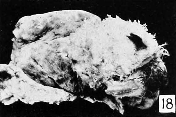
Fig. 18.
No. 1914 (Dr. G. C. McGorniick) is a fine, very characteristic mass, part of which is shown in figure 18. It is like Nos. 749 and 132.3, but very much larger, for in fluid it completely fills a 2-liter jar.
This specimen was said to have accompanied a living, 7-months fetus, having been expelled between the fetus and the placenta. Only a sniidl amount of clot, and what seems to be a small portion of placenta and membranes, accompanied it. Since the placenta was not saved it is impossible to say whether the mass resulted from partial degeneration of the placenta belonging to the living child, or whether it represented a degenerate twin placenta, which is rather unlikely but not imj)ossible, in view of the well- authenticated cases found in the literature.
This specimen is of interest not only for the numerous large, clear cysts, one of which measures 30X25 mm., which it contains, but because it accompanied the birth of a living child and because of the relative rareness of such a coincidence. In regard to the latter. Dr. McCormick added that in his experience of over 1 ,000 labors he had never before met this coincidence. The rareness of the specimen is emphasized still further by the statement of Professor Williams that such an instance has not been observed in a series of over 17,930 obstetrical cases treated by the department of obstetrics of the Johns Hopkins Medical School, as well as by the small series of such cases recorded in the literature.
Small chorionic vesicles, such as No. 2077 shown in natural size in figure 18, which attract no attention upon cursory inspection may, and often do, present the most exquisite picture of hydatiform degeneration when seen under a magnification of 3 to 20 diameters, as illustrated in figure 19. This is true especially if the examination is made with the binocular microscope. Since I have adopted this method of examination it has been possible to recognize instances of decidedly general and typical hydatiform degeneration in chorionic vesicles less than 2 cm. in size, with later confirmation of the diagnosis by a histologic examination. However, I have not been able to recognize very early stages merely by examination of the gross specimens, for gross recognition is possible only when portions of at least some of the villi have become sufficiently elliptical or globular to attract attention. Histologic recognition is possible far earlier than this, as shown in figure 20.
- Meyer Links: Plate 1 | Plate 2 | Plate 3 | Plate 4 | Plate 5 | Plate 6 | Contribution No.40 | Volume IX | Contributions to Embryology | Hydatidiform Mole | Tubal Pregnancy
| Historic Disclaimer - information about historic embryology pages |
|---|
 |
Reference
Meyer AW. Hydatiform degeneration in tubal and uterine pregnancy. (1920) Carnegie Instn. Wash. Publ., Contrib. Embryol., 40: 327- 364.
Cite this page: Hill, M.A. (2024, April 28) Embryology Meyer1920 fig18.jpg. Retrieved from https://embryology.med.unsw.edu.au/embryology/index.php/File:Meyer1920_fig18.jpg
- © Dr Mark Hill 2024, UNSW Embryology ISBN: 978 0 7334 2609 4 - UNSW CRICOS Provider Code No. 00098G
File history
Click on a date/time to view the file as it appeared at that time.
| Date/Time | Thumbnail | Dimensions | User | Comment | |
|---|---|---|---|---|---|
| current | 10:33, 8 April 2012 | | 570 × 379 (26 KB) | Z8600021 (talk | contribs) | ==Fig. 18.== Plate 3: Fig. 14 | Fig. 15 | Fig. 16 | Fig. 17 | Fig. 18 | |
You cannot overwrite this file.
File usage
The following page uses this file: