File:Meyer1920 fig09.jpg: Difference between revisions
No edit summary |
mNo edit summary |
||
| (2 intermediate revisions by the same user not shown) | |||
| Line 3: | Line 3: | ||
No. 323 (Dr. V. Van Williams) a large, firm, felt-like mass 120X90X65 mm., represented in figure 9. Very similar to, but a much larger specimen than in figure 8. | No. 323 (Dr. V. Van Williams) a large, firm, felt-like mass 120X90X65 mm., represented in figure 9. Very similar to, but a much larger specimen than in figure 8. | ||
The individual cysts, which vary from 1 to 20 mm., are packed together rather firmly, though a few large ones are free. The exterior of the specimen is formed by a thick layer of degenerate decidua and gives only a slight indication of its true nature upon closer inspection or upon examination of the cut surface. No fetal remnants were noticed, and microscopic examination shows | The individual cysts, which vary from 1 to 20 mm., are packed together rather firmly, though a few large ones are free. The exterior of the specimen is formed by a thick layer of degenerate decidua and gives only a slight indication of its true nature upon closer inspection or upon examination of the cut surface. No fetal remnants were noticed, and microscopic examination shows that the specimen is composed merely of a large hydatiform mass which was retained for a long tune and then aborted in toto with the surrounding decidua and exudate. | ||
Note: This same image appears in [[Book_-_Contributions_to_Embryology_Carnegie_Institution_No.56|Contributions Vol.12 No.56 (1921)]] as [[:File:Mall_Meyer1921_fig06.jpg|Figure 6]]. | |||
| Line 11: | Line 12: | ||
{{Meyer1920}} | {{Meyer1920}} | ||
[[Category:Hydatidiform Mole]] | |||
Latest revision as of 23:29, 11 May 2014
Fig. 8.
No. 323 (Dr. V. Van Williams) a large, firm, felt-like mass 120X90X65 mm., represented in figure 9. Very similar to, but a much larger specimen than in figure 8.
The individual cysts, which vary from 1 to 20 mm., are packed together rather firmly, though a few large ones are free. The exterior of the specimen is formed by a thick layer of degenerate decidua and gives only a slight indication of its true nature upon closer inspection or upon examination of the cut surface. No fetal remnants were noticed, and microscopic examination shows that the specimen is composed merely of a large hydatiform mass which was retained for a long tune and then aborted in toto with the surrounding decidua and exudate.
Note: This same image appears in Contributions Vol.12 No.56 (1921) as Figure 6.
Plate 2: Fig. 8 | Fig. 9 | Fig. 10 | Fig. 11 | Fig. 12 | Fig. 13
- Meyer Links: Plate 1 | Plate 2 | Plate 3 | Plate 4 | Plate 5 | Plate 6 | Contribution No.40 | Volume IX | Contributions to Embryology | Hydatidiform Mole | Tubal Pregnancy
| Historic Disclaimer - information about historic embryology pages |
|---|
 |
Reference
Meyer AW. Hydatiform degeneration in tubal and uterine pregnancy. (1920) Carnegie Instn. Wash. Publ., Contrib. Embryol., 40: 327- 364.
Cite this page: Hill, M.A. (2024, May 19) Embryology Meyer1920 fig09.jpg. Retrieved from https://embryology.med.unsw.edu.au/embryology/index.php/File:Meyer1920_fig09.jpg
- © Dr Mark Hill 2024, UNSW Embryology ISBN: 978 0 7334 2609 4 - UNSW CRICOS Provider Code No. 00098G
File history
Click on a date/time to view the file as it appeared at that time.
| Date/Time | Thumbnail | Dimensions | User | Comment | |
|---|---|---|---|---|---|
| current | 09:57, 8 April 2012 | 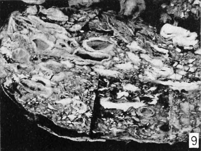 | 794 × 597 (68 KB) | Z8600021 (talk | contribs) | ==Fig. 8.== Plate 2: Fig. 8 | Fig. 9 | Fig. 10 | Fig. 11 | Fig. 12 | [[ |
You cannot overwrite this file.
File usage
The following page uses this file: