File:Meyer1920 fig04.jpg
Original file (491 × 648 pixels, file size: 32 KB, MIME type: image/jpeg)
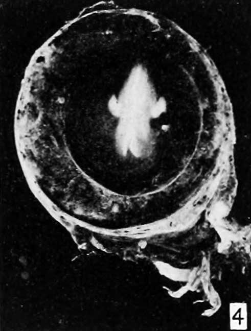
Fig. 4.
The wall of the tube is quite thin, as figure 4 shows, but the implantation is fairly well preserved around the whole perimeter of the specimen.
The mucosa is destroyed throughout the greater extent of the section and the trophoblast is abundant, except in one rather degenerate and hemorrhagic area. The chorionic membrane is thin but contains some vessels distended with blood. The stroma of many of the villi also contains vessels filled with blood, but the vessels in many others are very evidently in degeneration.
The syncytium is scanty and many of the villi are very plainly hydatiform, as seen in figures 5 and 6.
- Meyer Links: Plate 1 | Plate 2 | Plate 3 | Plate 4 | Plate 5 | Plate 6 | Contribution No.40 | Volume IX | Contributions to Embryology | Hydatidiform Mole | Tubal Pregnancy
| Historic Disclaimer - information about historic embryology pages |
|---|
 |
Reference
Meyer AW. Hydatiform degeneration in tubal and uterine pregnancy. (1920) Carnegie Instn. Wash. Publ., Contrib. Embryol., 40: 327- 364.
Cite this page: Hill, M.A. (2024, April 27) Embryology Meyer1920 fig04.jpg. Retrieved from https://embryology.med.unsw.edu.au/embryology/index.php/File:Meyer1920_fig04.jpg
- © Dr Mark Hill 2024, UNSW Embryology ISBN: 978 0 7334 2609 4 - UNSW CRICOS Provider Code No. 00098G
File history
Click on a date/time to view the file as it appeared at that time.
| Date/Time | Thumbnail | Dimensions | User | Comment | |
|---|---|---|---|---|---|
| current | 09:21, 8 April 2012 | | 491 × 648 (32 KB) | Z8600021 (talk | contribs) | ==Fig. 1.== Plate 1: Fig. 1 | Fig. 2 | Fig. 3 | Fig. 4 | Fig. 5 | [[:Fi |
You cannot overwrite this file.
File usage
The following 2 pages use this file: