File:Meyer1920 fig03.jpg
Meyer1920_fig03.jpg (450 × 600 pixels, file size: 20 KB, MIME type: image/jpeg)
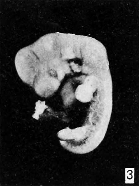
Fig. 3.
Another very interesting specimen of tubal implantation is No. 1771, received from Dr. H. M. N. Wynne, of the Johns Hopkins Hospital. The menstrual age of this specimen is 49 days, but its anatomic age, as based upon length according to Dr. Streeter's curve (unpublished), is 37 days, thus showing a discrepancy between the menstrual and anatomic ages of 12 days. The embryonic length is only 12.5 mm., although with a menstrual age of 49 days it should be at least 18 mm. Upon examination, Dr. Streeter found the chorionic vesicle to contain a good deal of magma, some of which still was adherent to the embryo, as figure 3 shows. As has been repeatedly emphasized in the literature, the presence of this coagulum in itself probably indicates that the embryo died some time previously.
Plate 1: Fig. 1 | Fig. 2 | Fig. 3 | Fig. 4 | Fig. 5 | Fig. 6 | Fig. 7
- Meyer Links: Plate 1 | Plate 2 | Plate 3 | Plate 4 | Plate 5 | Plate 6 | Contribution No.40 | Volume IX | Contributions to Embryology | Hydatidiform Mole | Tubal Pregnancy
| Historic Disclaimer - information about historic embryology pages |
|---|
 |
Reference
Meyer AW. Hydatiform degeneration in tubal and uterine pregnancy. (1920) Carnegie Instn. Wash. Publ., Contrib. Embryol., 40: 327- 364.
Cite this page: Hill, M.A. (2024, April 28) Embryology Meyer1920 fig03.jpg. Retrieved from https://embryology.med.unsw.edu.au/embryology/index.php/File:Meyer1920_fig03.jpg
- © Dr Mark Hill 2024, UNSW Embryology ISBN: 978 0 7334 2609 4 - UNSW CRICOS Provider Code No. 00098G
File history
Click on a date/time to view the file as it appeared at that time.
| Date/Time | Thumbnail | Dimensions | User | Comment | |
|---|---|---|---|---|---|
| current | 09:21, 8 April 2012 | | 450 × 600 (20 KB) | Z8600021 (talk | contribs) | ==Fig. 1.== Plate 1: Fig. 1 | Fig. 2 | Fig. 3 | Fig. 4 | Fig. 5 | [[:Fi |
You cannot overwrite this file.
File usage
The following 2 pages use this file: